Visible reamer structure under microscope

A reamer and endoscope technology, applied in the field of visible reamer structure under the microscope, can solve the problems of inconvenient reamer application force, prolonged operation time, and cumbersome expansion steps, so as to improve accuracy, ensure safety, reduce The effect of times

Pending Publication Date: 2018-06-19
ZHUHAI WEIERKANG BIOTECH
View PDF5 Cites 3 Cited by
  • Summary
  • Abstract
  • Description
  • Claims
  • Application Information

AI Technical Summary

Problems solved by technology

[0002] In the existing medical technology, the puncture needle is mostly used as a guide, but the puncture needle does not have a fixed effect, and it is not convenient for the operator to use the force of the reamer, and the target site cannot be accurately excised
At the same time, the positioning of the reamer mostly adopts the method of X-ray positioning. The X-ray positioning is not accurate enough to ensure the safety of the operation.
In addition, in the prior art, a group of Tom needles are used to continuously change the diameter and expand step by step. The expansion steps are cumbersome and require multiple X-ray positioning assistance, which greatly increases the damage of doctors and patients exposed to X-rays and prolongs the operation time.

Method used


Image

  • Visible reamer structure under microscope
  • Visible reamer structure under microscope
  • Visible reamer structure under microscope

Examples


Embodiment Construction

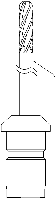
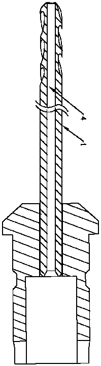
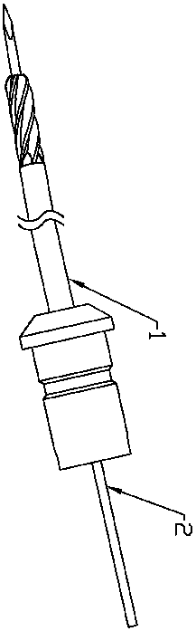
[0013] Such as Figure 1 to Figure 5 As shown, in the present embodiment, the present invention includes a reamer 1, a Kirschner wire 2, an operation endoscope structure 3, and a manual or electric tool for fixing the Kirschner wire into bone tissue, and the shaft of the reamer 1 A hollow channel 4 is provided, and the Kirschner wire 2 passes through the hollow channel 4. The surgical endoscope structure 3 includes a body 5 and a working channel 6 connected to the body 5. The hinge The knife 1 penetrates from one end of the working channel 6 and passes out from the other end of the working channel 6 . In this embodiment, an imaging module and a light source are also arranged in the working channel 6 .

[0014] In this embodiment, the cutter head of the reamer 1 is a full-face sawtooth, and the full-face sawtooth includes side sawtooth and top surface sawtooth. The cutter head of the reamer 1 in the present invention is fully serrated, which can effectively cut off the front ...


PUM


Abstract

The invention discloses a visible reamer structure under a microscope, is applied to the technical field of the visible reamer structure under the microscope, and aims at achieving the technical purpose of providing the visible reamer structure which can operate a reamer in a visible environment and is easy and fast to operate, high in operation efficiency and higher in safety under the microscope. The visible reamer structure comprises the reamer, a Kirschner wire, an operation endoscope structure and a manual or electric tool pushing the Kirschner wire into the synostosis tissue to be fixed,a hollow channel is formed in the axial direction of the reamer, an annular saw base is arranged at the tail end of the reamer and can be connected with a T-shaped handle, the Kirschner wire penetrates through the hollow channel, the operation endoscope structure comprises a body and a working channel connected with the body, and the reamer penetrates into one end of the working channel and penetrates out of the other end of the working channel.

Description

technical field [0001] The invention relates to a reamer, in particular to a structure of a reamer visible under a microscope. Background technique [0002] In the existing medical technology, the puncture needle is mostly used for guidance, but the puncture needle does not have a fixing effect, and it is not convenient for the operator to use the force of the reamer, and the target site cannot be accurately excised. At the same time, the positioning of the reamer mostly adopts the method of X-ray positioning, but the X-ray positioning is not accurate enough, and it is difficult to ensure the safety of the operation. In addition, in the prior art, a group of Tom needles are continuously changed in diameter and expanded step by step. The expansion steps are cumbersome and require multiple X-ray positioning assistance, which greatly increases the damage of doctors and patients exposed to X-rays and prolongs the operation time. Contents of the invention [0003] The technica...

Claims


Application Information

Patent Timeline
Patent Type & Authority Applications(China)
IPC IPC(8): A61B17/00A61B17/14A61B17/88A61B17/3205
CPCA61B17/00234A61B17/14A61B17/3205A61B17/88A61B2017/564
Inventor 胡善云卢宏浩
Owner ZHUHAI WEIERKANG BIOTECH
Why Eureka
Try Eureka