Color normalization method for HE staining pathological images

A pathological image and normalization technology, applied in the field of image processing, can solve the problems of slowing down the normalization process, large computational load of cell nucleus detection and segmentation algorithms, and inability to obtain the normalization effect.

Active Publication Date: 2018-06-15
BEIHANG UNIV
View PDF5 Cites 5 Cited by
  • Summary
  • Abstract
  • Description
  • Claims
  • Application Information

AI Technical Summary

Problems solved by technology

However, the effect of this type of method depends on the nucleus detection and segmentation algorithm. Once the nucleus detection algorithm fails, a reasonable normalization effect cannot be obtained, and the calculation of the nucleus detection and segmentation algorithm is large, which slows down the entire normalization. process

Method used


Image

  • Color normalization method for HE staining pathological images
  • Color normalization method for HE staining pathological images
  • Color normalization method for HE staining pathological images

Examples


Embodiment Construction

[0057] The technical solutions in the embodiments of the present invention will be clearly and completely described below with reference to the accompanying drawings in the embodiments of the present invention. Obviously, the described embodiments are only a part of the embodiments of the present invention, but not all of the embodiments. Based on the embodiments of the present invention, all other embodiments obtained by those of ordinary skill in the art without creative efforts shall fall within the protection scope of the present invention.

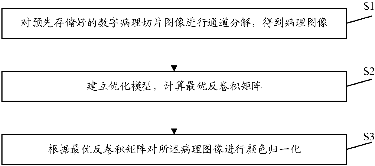
[0058] see attached figure 1 , the embodiment of the present invention discloses a color normalization method for HE staining pathological images, including:

[0059] S1: Perform channel decomposition on the pre-stored digital pathological slice image to obtain the pathological image I(x, y);

[0060]

[0061] Among them, I R (x,y), I G (x,y), I B (x, y) represent the pixel values ​​of the pathological image in the three color ...


PUM


Abstract

The invention discloses a color normalization method for HE staining pathological images. The method comprises the following steps: a step of subjecting pathological slice images to channel decomposition operation and obtaining pathological images, a step of calculating an optimal deconvolution matrix via establishment of an optimization model, and a step of using the optimal deconvolution matrixto normalizing the pathological images; the color of the pathological images under different staining conditions tends to be uniform after normalization, and fast calculation speed is realized.

Description

technical field [0001] The invention relates to the technical field of image processing, in particular to a color normalization method for HE staining pathological images. Background technique [0002] The digital pathological full slide is to convert the pathological slide into a high-magnification large-scale digital image that can be displayed, transmitted and processed by a computer through a special scanning imaging system. With the continuous maturity of digital pathological image imaging technology, computer-aided diagnosis methods based on digital pathological images have developed rapidly. With the deepening of research, the auxiliary diagnosis method has been developed in the direction of pathological whole slide image (WSI) adapted to multi-staining platform and scanning platform. However, affected by the ratio of dyes, dyeing platform, and imaging platform, the color of WSI from different platforms varies greatly. This difference greatly affects the accuracy of...

Claims


Application Information

Patent Timeline
Patent Type & Authority Applications(China)
IPC IPC(8): G16H30/40G06T7/90
CPCG06T2207/10024G06T2207/10056G06T2207/30024G06T7/90
Inventor 姜志国张浩鹏郑钰山谢凤英
Owner BEIHANG UNIV
Why Eureka
Try Eureka