Preparation method of near infrared fluorescence imaging micromolecule anticancer nano-drug

A nano-drug and fluorescence imaging technology, applied in the field of biomedicine, can solve the problems of unclear mechanism of action, complex nano-system, unclear metabolism, etc., and achieve the effect of improving photostability, simple preparation process, and eliminating clinical safety problems.

Active Publication Date: 2018-03-20
FUZHOU UNIV
View PDF3 Cites 9 Cited by
  • Summary
  • Abstract
  • Description
  • Claims
  • Application Information

AI Technical Summary

Problems solved by technology

[0008] The purpose of the present invention is to prepare a method for preparing near-infrared fluorescence imaging small molecule anticancer nano-medicines through the interaction of UA, PTX and photosensitizer ICG with anticancer activity through π-π stacking electrostatic force, to solv

Method used


Image

  • Preparation method of near infrared fluorescence imaging micromolecule anticancer nano-drug
  • Preparation method of near infrared fluorescence imaging micromolecule anticancer nano-drug
  • Preparation method of near infrared fluorescence imaging micromolecule anticancer nano-drug

Examples


Embodiment 1

[0045] The preparation method of ursolic acid nano micelles

[0046] Accurately weigh 0.00456g of UA powder, dissolve in 1ml of methanol, ultrasonically dissolve, and configure a 10 mM solution; take different volumes of methanol solution, and add dropwise to the solution containing 2 mL of secondary water (double distilled water) during the stirring process. ) (Note: Stir at high speed during the dropping process, and the dropping time is 30s), at this time, the concentration of UA in the solution is 31.25 μM-1000 μM, and then stir for 5 min to obtain UA nanomicelles;

[0047] The average particle size and PID of UA nanomicelles with different concentrations prepared in this example are shown in Table 1.

[0048] Table 1

[0049]

Embodiment 2

[0051] Accurately weigh 0.00853g of PTX powder, dissolve it in 1ml of methanol, and ultrasonically dissolve it to form a 10mM solution; take different volumes of methanol solution, and add it dropwise to 2ml of secondary water during the stirring process (note: the dropping process Medium-high speed stirring, dropping time is 30s), at this time, the concentration of PTX in the solution is 31.25μM-1000μM, and then stirring for 5min to obtain PTX nanomicelle;

[0052] The average particle size and PID of PTX nanomicelles with different concentrations prepared in this example are shown in Table 2.

[0053] Table 2

[0054]

Embodiment 3

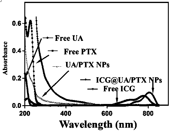
[0056] Accurately weigh 0.00456g of UA powder and 0.00853g of PTX powder, dissolve them in 1ml of methanol, configure them into a 10mM solution, and ultrasonically dissolve them; take different volumes of ursolic acid in methanol and different volumes of paclitaxel in methanol, mix them, and stir During the process, add dropwise to secondary water containing 2ml (note: stir at high speed during the dropping process, and the dropping time is 30s), and then stir for 5min to obtain UA / PTX nanomicelles with different molecular molar ratios;

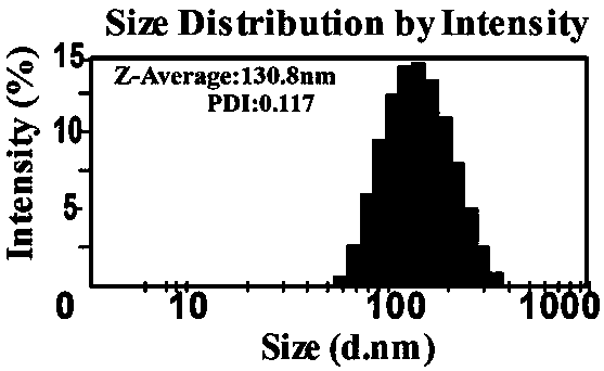
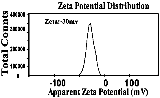
[0057] Table 3 shows the average particle size, PID and potential of the UA / PTX nanomicelles formed by different molecular molar ratios prepared in this example.

[0058] table 3

[0059]


PUM

PropertyMeasurementUnit
Particle sizeaaaaaaaaaa

Abstract

The invention provides a preparation method of a near infrared fluorescence imaging micromolecule anticancer nano-drug. The method comprises the steps of using hydrophobic drugs UA (ursolic acid) andPTX (paclitaxel) for preparing into a nano-micelle, adopting the nano-micelle as a carrier, and co-assembling the carrier and a hydrophilic ICG to obtain the nano-drug. The invention aims to provide the preparation method for preparing the near infrared fluorescence imaging micromolecule anticancer nano-drug through interaction of pi-pi accumulation electrostatic forces of the UA and the PTX withantitumor activity and the photosensitizer ICG, so that the problems that in the prior art, an artificially synthesized carrier nanometer system is complex in system, difficult in quality control, undefined in action mechanism, unclear in metabolism and the like are solved, and the aim of treating tumors through the synergism of phototherapy and chemotherapy is achieved.

Description

technical field [0001] The invention relates to the technical field of biomedicine, in particular to paclitaxel with anticancer activity, ursolic acid with anticancer activity and indocyanine green, a photosensitizer, co-assembled into nano micelles, and prepared into a near-infrared A method for fluorescence imaging of small molecule anticancer nanomedicines. Background technique [0002] Tumor is one of the three major diseases that seriously threaten human health today, but the overall morbidity and mortality of cancer are still rising worldwide. very important. Traditional chemotherapy methods have disadvantages such as high toxicity and side effects, which cause great physical and mental damage to patients. With the rapid development of science and technology, the research of antineoplastic drugs is being paid more and more attention by people. The development of new materials and new drugs for targeted therapy of tumors is an urgent problem to be solved. [0003] N...

Claims


Application Information

Patent Timeline
IPC IPC(8): A61K31/56A61K31/337A61K9/107A61K47/28A61K41/00A61P35/00
CPCA61K9/1075A61K31/337A61K31/56A61K41/0057A61K47/28A61K2300/00
Inventor 邵敬伟郭燕林娟芳许爱笑
Owner FUZHOU UNIV
Why Eureka
Try Eureka