A biomarker for esophageal cancer and its application
A biomarker, esophageal cancer technology, applied in the field of esophageal cancer biomarkers
- Summary
- Abstract
- Description
- Claims
- Application Information
AI Technical Summary
Problems solved by technology
Method used
Image
Examples
Embodiment 1
[0050] collect samples
[0051]Collect 100 cases of surgically resected esophageal cancer specimens and 80 cases of normal tissues, as well as 40 cases of primary esophageal cancer metastasis tissues purchased from Shanghai Xinchao Biotechnology Co., Ltd.
[0052] tissue microarray
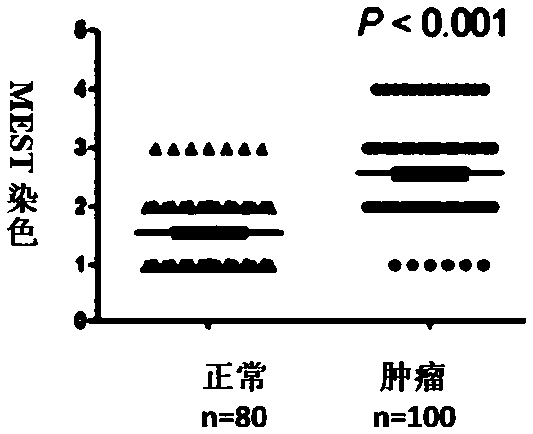
[0053] Tissue microarray (TMA) (Biomax, Rockville, MD) immunohistochemical method was used to analyze the expression of MEST on 100 cases of esophageal cancer tissues and 80 cases of normal tissues to determine the correlation between the upregulation of MEST expression and the occurrence and development of esophageal cancer and perform MEST immunohistochemical analysis on another tissue microarray (TMA) of 40 primary esophageal cancer metastatic tissues purchased by Shanghai Xinchao Biotechnology Co., Ltd. to compare the expression of MEST in primary lesions and metastatic lesions Then, the correlation between MEST expression level and tumor metastasis was determined.
[0054] Immunohistochemis...
Embodiment 2
[0057] collect samples
[0058] Sera from 100 patients and 100 healthy controls were collected from Jinan University and Shantou University.
[0059] ELISA detection
[0060] The expression of MEST in the serum of 100 patients with esophageal cancer and 100 healthy controls was detected by enzyme-linked immunosorbent assay (ELISA) to determine the correlation between the upregulation of MEST expression and esophageal squamous cell carcinoma.
Embodiment 3
[0061] Embodiment 3 result analysis
[0062] 1. MEST expression
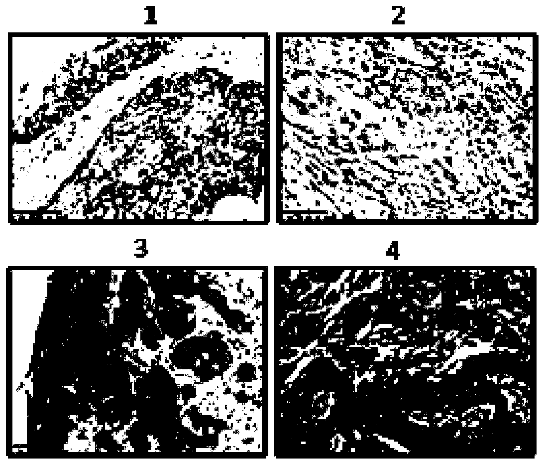
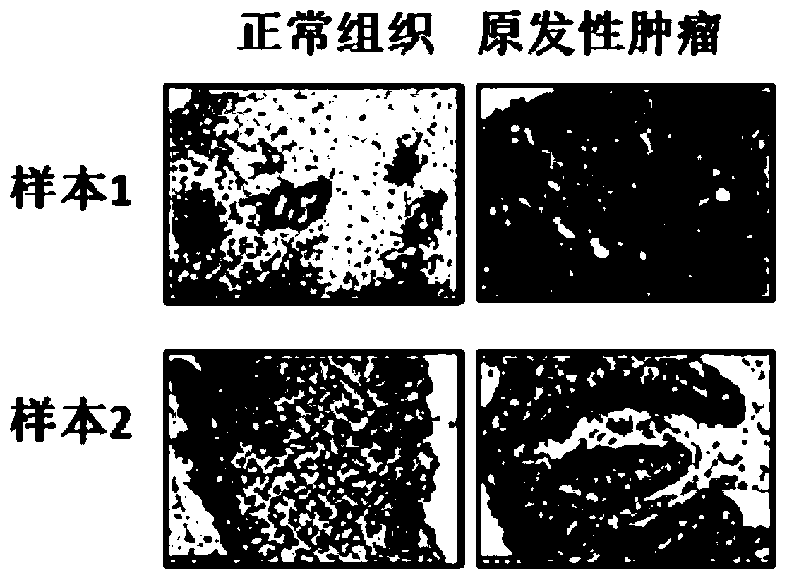
[0063] In order to study the clinical significance of MEST in human tumors, the expression level of MEST was detected by immunohistochemistry, and the expression level of MEST in 100 cases of esophageal cancer tissues and 80 cases of normal tissues was analyzed by tissue microarray method. The dyeing results are scored, and the scoring rules are as follows: figure 1 As shown, the staining intensity of TMA chip scanning: divided into four grades (1-4), 1 point represents negative, 2 points represent weak positive, 3 points represent medium positive, 4 points represent strong positive. Two pathological methods were used to test and independently score the immunostaining degree of the sections. Samples with a score of 1 to 2 were considered low expression, and samples with a score of 3 to 4 were considered high expression; normal tissue and esophageal cancer tissue The staining results and analysis charts are sho...
PUM
 Abstract
Description
Claims
Application Information
 - Generate Ideas
- Intellectual Property
- Life Sciences
- Materials
- Tech Scout
- Unparalleled Data Quality
- Higher Quality Content
- 60% Fewer Hallucinations
Browse by: Latest US Patents, China's latest patents, Technical Efficacy Thesaurus, Application Domain, Technology Topic, Popular Technical Reports.
© 2025 PatSnap. All rights reserved.Legal|Privacy policy|Modern Slavery Act Transparency Statement|Sitemap|About US| Contact US: help@patsnap.com


