Mouse Shank 3 gene cRNA probe and in-situ hybridization color development method

An in situ hybridization, mouse technology, applied in the field of biomedicine, can solve the problems that the color development effect needs to be improved, the composition is complex, the cRNA probe and the fluorescent in situ hybridization color development method has not been reported, and the processing time can be optimized. , long storage time, economical and convenient effect

Active Publication Date: 2018-01-12
FOURTH MILITARY MEDICAL UNIVERSITY
View PDF3 Cites 3 Cited by
  • Summary
  • Abstract
  • Description
  • Claims
  • Application Information

AI Technical Summary

Problems solved by technology

However, screening cRNA probes with both hybridization specificity and chromogenic sensitivity for the Shank family, especially the Shank3 gene, is still a difficult problem in the field
Although the use of reagents with the function of amplifying the color development effect is more conducive to color development, there is no amplification system for cRNA probe in situ hybridization color development suitable for the Shank family, especially Shank3, which also leads to the corresponding An important reason why cRNA probes have not been obtained
In addition, the existing cRNA probe-based chromogenic methods have disadvantages such as complex composition of chromogenic reagents (such as amplification reagents, hybridization solutions), and at the same time, the chromogenic effect still needs to be improved
[0004] So far, there are no reports on cRNA probes and fluorescent in situ hybridization methods for Shank3 gene

Method used


Image

  • Mouse Shank 3 gene cRNA probe and in-situ hybridization color development method
  • Mouse Shank 3 gene cRNA probe and in-situ hybridization color development method
  • Mouse Shank 3 gene cRNA probe and in-situ hybridization color development method

Examples


Embodiment Construction

[0029] The present invention will be described in further detail below in conjunction with the accompanying drawings and embodiments. The examples are used to explain the present invention, not to limit the present invention.

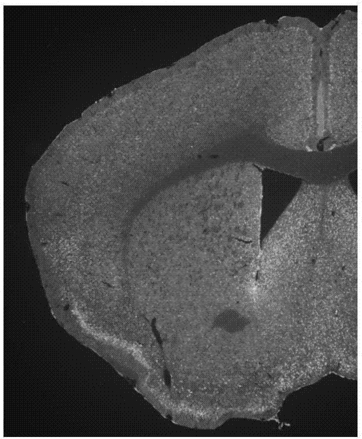
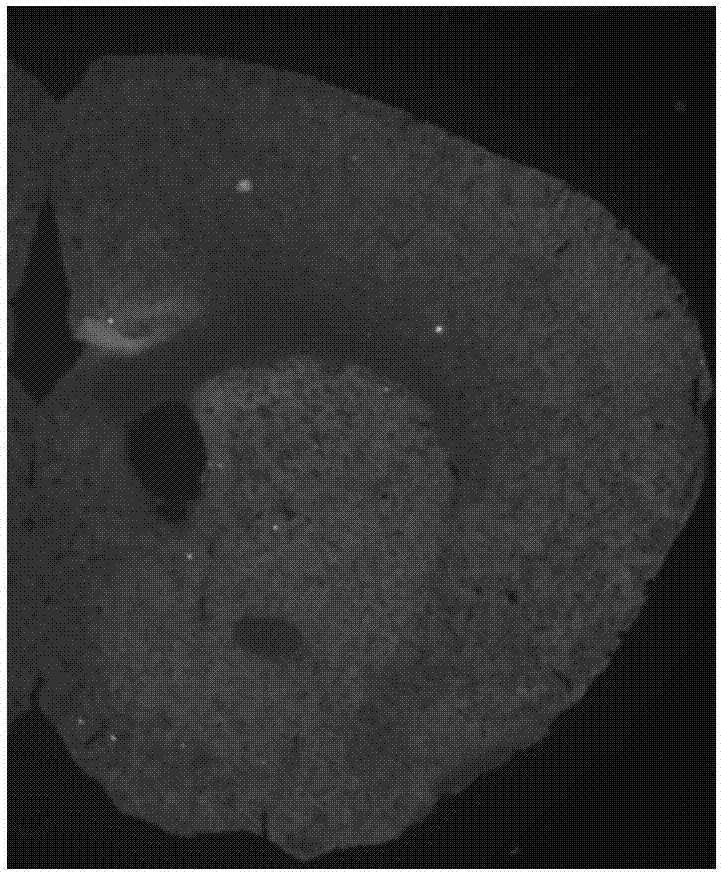
[0030] The invention constructs the cRNA probe of the mouse Shank3 gene, and clearly shows the expression of the mouse Shank3 gene in normal mouse brain tissue by means of fluorescence in situ hybridization, providing an experimental basis for further double labeling.

[0031] 1. Shank 3 gene cRNA probe preparation

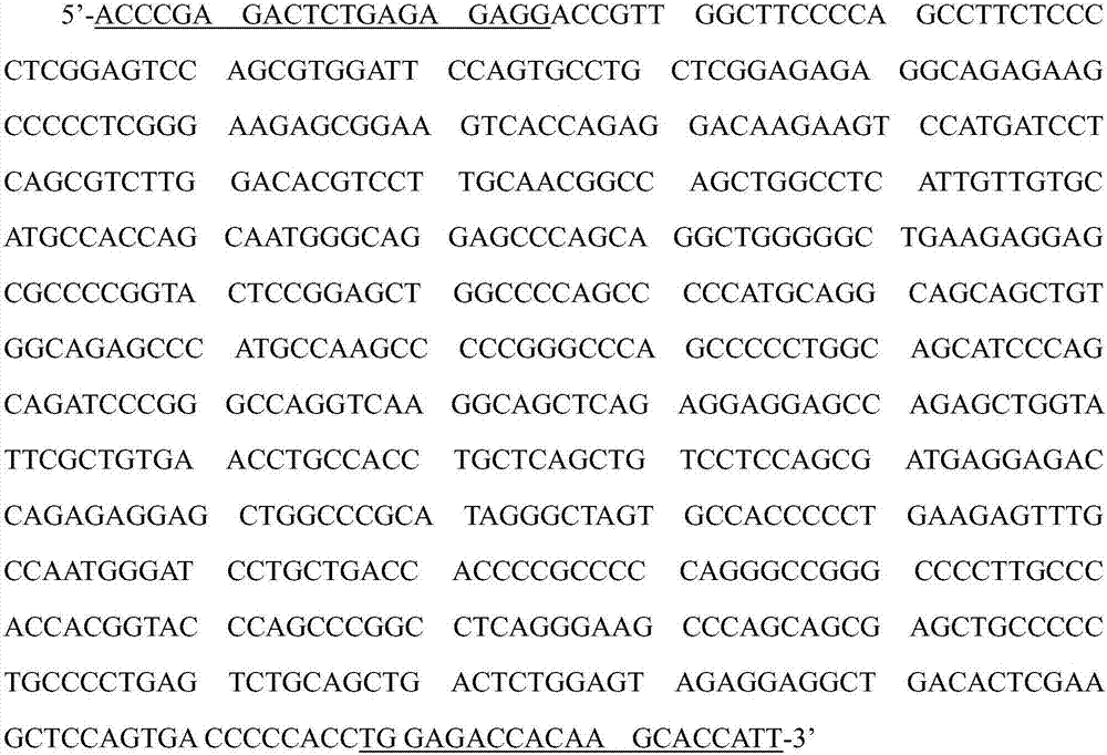
[0032] 1. According to the reference sequence (NM_021423.3) of the mouse Shank 3 gene mRNA in the Gene bank, blastn was used to select relatively specific fragments, and in combination with the fluorescence in situ hybridization method proposed by the present invention, the Shank3 cRNA was finally determined. Specific fragments (nucleotides 3465-4108; GenBank accession No. NM_021423.3).

[0033] 2. Using the primer-blast tool for this fra...


PUM


Abstract

The invention provides a mouse Shank 3 gene cRNA probe and an in-situ hybridization color development method. The cRNA probe comprises a nucleotide sequence with complementary sites 3465-4108 of a reference sequence NM_021423.3 of mouse Shank 3 gene mRNA. The in-situ hybridization color development method comprises the following steps: putting a mouse brain tissue sample in a hybridization solution; after hybridization, rinsing, closing and combining with an antibody; amplifying with TSA-biotin and performing fluorescent color development. The cRNA probe provided by the invention has high specificity on the Shank 3 gene, can efficiently finish recognition of the Shank 3 gene in the brain tissues of different individuals, can ensure the specificity and high efficiency of in-situ hybridization color development of the mouse Shank 3 gene, realizes a better color development effect and can be combined with other hybridization or immunofluorescent staining.

Description

technical field [0001] The invention belongs to the field of biomedicine, and in particular relates to a Shank 3 gene cRNA probe and a fluorescence in situ hybridization color development method thereof. Background technique [0002] The Shank family is a newly discovered scaffold protein, which can interact with various proteins in cells through different structural domains and play multiple roles. Studies have shown that the Shank3 gene is closely related to the onset of autism, and mutations in the Shank3 gene can lead to autism-like behaviors. Studies such as protein interaction have shown that Shank3 protein may interact with various intracellular proteins through different structural domains, and ultimately affect the structure and function of the cytoskeleton and synapses. However, the morphological evidence in this regard is obviously insufficient, mainly due to the fact that none of the commercially available antibodies can clearly show the intracellular localizati...

Claims


Application Information

Patent Timeline
IPC IPC(8): C12Q1/6841C12Q1/6804C12Q1/6883
Inventor 陈晶郭保霖蔡国洪吴菲菲武胜昔
Owner FOURTH MILITARY MEDICAL UNIVERSITY
Why Eureka
Try Eureka