A method and device for segmenting lung lobes based on CT images

A technology of CT images and lung lobes, which is applied in the field of image processing, can solve problems such as no lung lobes segmentation, and achieve the effect of saving time

Active Publication Date: 2020-01-07
NEUSOFT MEDICAL SYST CO LTD
View PDF3 Cites 0 Cited by
  • Summary
  • Abstract
  • Description
  • Claims
  • Application Information

AI Technical Summary

Problems solved by technology

[0004] At present, chest solutions in the prior art mainly focus on whole lung segmentation, tracheal segmentation, angiographic imaging and emphysema analysis, and do not involve detailed segmentation of lung lobes

Method used


Image

  • A method and device for segmenting lung lobes based on CT images
  • A method and device for segmenting lung lobes based on CT images
  • A method and device for segmenting lung lobes based on CT images

Examples


Embodiment 1

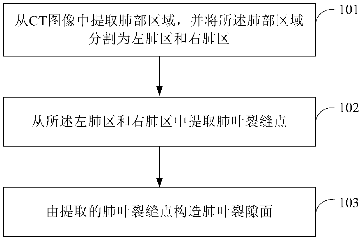
[0067] see figure 1 , which is a flow chart of Embodiment 1 of the method for segmenting lung lobes based on CT images provided by the present invention.

[0068] The method for the segmentation of lung lobes based on CT images provided in this embodiment comprises the following steps:

[0069] Step 101: extract a lung area from a CT image, and segment the lung area into a left lung area and a right lung area.

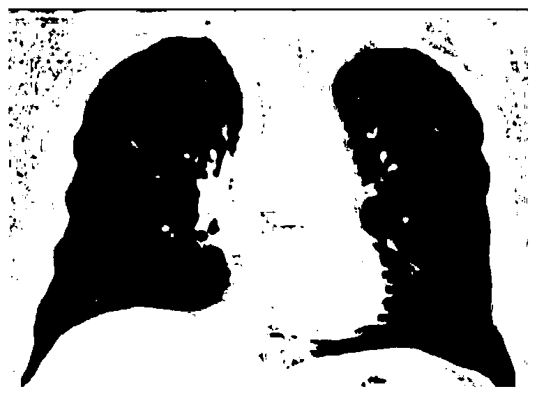
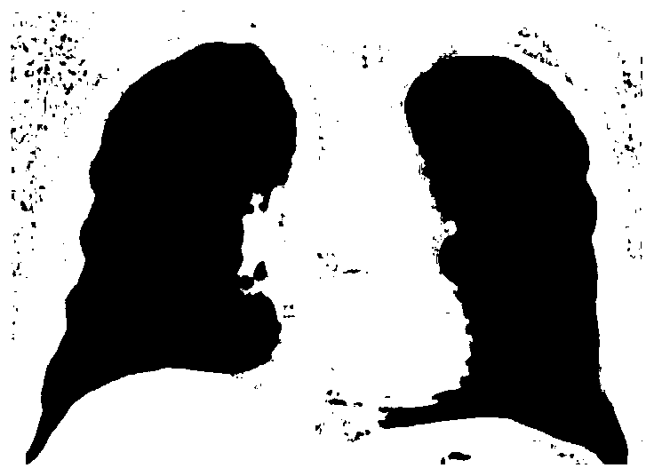
[0070] It should be noted that, generally, when taking a CT image of the lungs, the image of the chest is directly taken. Therefore, it is necessary to extract the lung area from the chest CT image and determine the boundary between the left lung area and the right lung area. After determining the area to which the left and right lungs belong, the lung lobe segmentation can be performed in the left lung area and the right lung area. Generally, the image of the lung area extracted from the CT image, such as figure 2 shown. The left lung area includes 2 lobes, and th...

Embodiment 2

[0077] The initial location of each lobe is connected to a bronchi. In accordance with this physiological characteristic, this embodiment divides the airway into two levels, the first level is the trachea, and the second level is the bronchi. The end of the bronchi is used to find the initial position of each lung lobe. The following describes the lung trachea in the CT image Segmentation is performed to determine the initial location of the lung lobes.

[0078] see Figure 6 , which is a flow chart of Embodiment 2 of the method for segmenting lung lobes based on CT images provided by the present invention.

[0079] The method for lung lobe segmentation provided in this embodiment includes:

[0080] Step 201: Extract lung regions from CT images.

[0081] Step 202: Segment the lung region into a left lung region and a right lung region.

[0082] Step 203: Segment the trachea and bronchi from the CT image of the lung region.

[0083] Step 202 and step 203 have no sequence r...

Embodiment 3

[0095] Since there are many image points in the acquired CT image, some of the image points may be interference points caused by certain factors in the process of obtaining the CT image, and some points may be points on other organs of the human body. Such as crack local bright spot, vessel wall signal point, etc. These image points mentioned above will all produce interference during the process of extracting the fissure points of the lung lobe. Therefore, in order to extract the fissure points of the lung lobe more accurately, these interference points need to be suppressed.

[0096] see Figure 7 , which is a flow chart of Embodiment 3 of the method for segmenting lung lobes based on CT images provided by the present invention.

[0097] The method for segmenting lung lobes based on CT images provided in this embodiment includes:

[0098] Step 301: extract a lung area from a CT image, and segment the lung area into a left lung area and a right lung area.

[0099] Step 302...


PUM


Abstract

The present invention provides a CT image-based lung lobe segmentation method and device. The method includes the following steps that: a lung region is extracted from a CT image, the lung region is divided into a left lung region and a right lung region; lung lobe crack points are extracted from the left lung region and the right lung region; and a lung lobe crack surface is constructed on the basis of the lung lobe crack points. According to the CT image-based lung lobe segmentation method and device of the invention, the lung region is extracted from the CT image, and the lung region is divided into the left lung region and the right lung region, wherein the left lung region contains two lung lobes, and the right lung region contains three lung lobes; the lung lobe crack points are extracted from the left lung region and the right lung region; and the lung lobe crack surface is constructed on the basis of the lung lobe crack points. With the method and device of the invention adopted, lung lobe segmentation can be automatically performed; after the lung lobe segmentation is performed through the method, if five clear lung lobes cannot be obtained, it can be indicated that physiological abnormalities exist in lungs, or tumors exists around the in the lungs. Since the method can automatically segment the lungs into five lung lobes, if the method fails to obtain five lung lobes through segmentation, it is indicated that the abnormalities exist in the lungs, and therefore, a lot of time can be saved for doctors; and the doctors do not have to spend a lot of time manually analyzing CT images to find suspicious areas.

Description

technical field [0001] The present invention relates to the technical field of image processing, in particular to a lung lobe segmentation method and device based on CT images. Background technique [0002] Lung disease has gradually become the most common disease that threatens human life and health. Among them, lung cancer has become the biggest threat among cancers because of its difficulty in detection, difficulty in treatment after discovery, and high mortality. When patients suffer from lung cancer with significant symptoms, they have basically missed the best period of diagnosis and treatment, so early diagnosis and treatment of lung cancer are very important. At present, low-dose spiral CT is mainly used in clinical diagnosis and screening of pulmonary nodules. The emergence and improvement of the computer-aided diagnosis system for the diagnosis of lung cancer is the most important thing in the development of contemporary medical care. [0003] Since the developme...

Claims


Application Information

Patent Timeline
Patent Type & Authority Patents(China)
IPC IPC(8): G06T7/10
CPCG06T7/10G06T2207/10081G06T2207/30061
Inventor 陈磊曲凯晨康雁吕晓凤
Owner NEUSOFT MEDICAL SYST CO LTD
Why Eureka
Try Eureka