Biological chip for carrying out screening positioning and detection on rare cells in blood
A rare cell and biochip technology, applied in the field of biochips for screening, locating and detecting rare cells in blood, can solve problems such as damage to tumor cells, identification process interference, escape, etc., and achieve efficient capture, rapid analysis, and convenient follow-up detection. Effect
- Summary
- Abstract
- Description
- Claims
- Application Information
AI Technical Summary
Problems solved by technology
Method used
Image
Examples
Embodiment 1
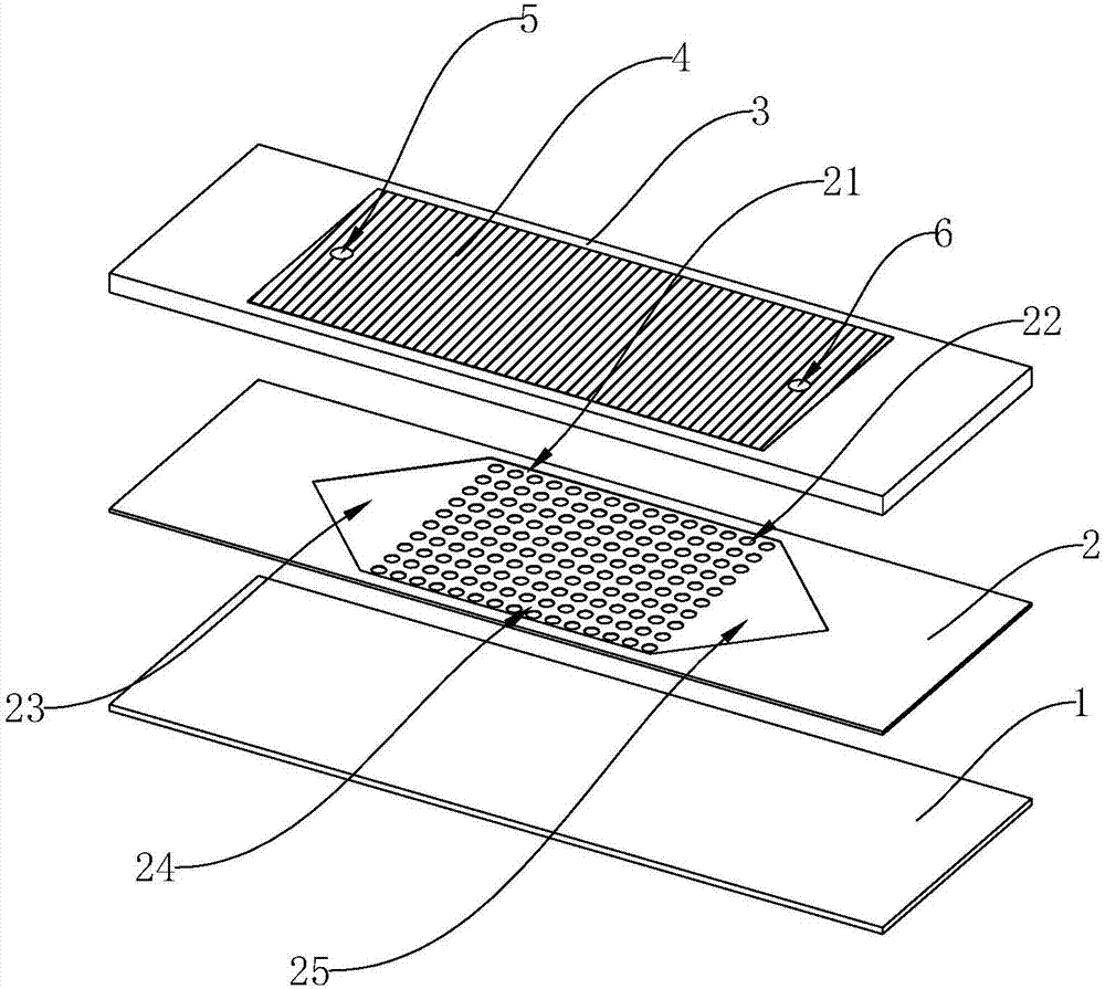
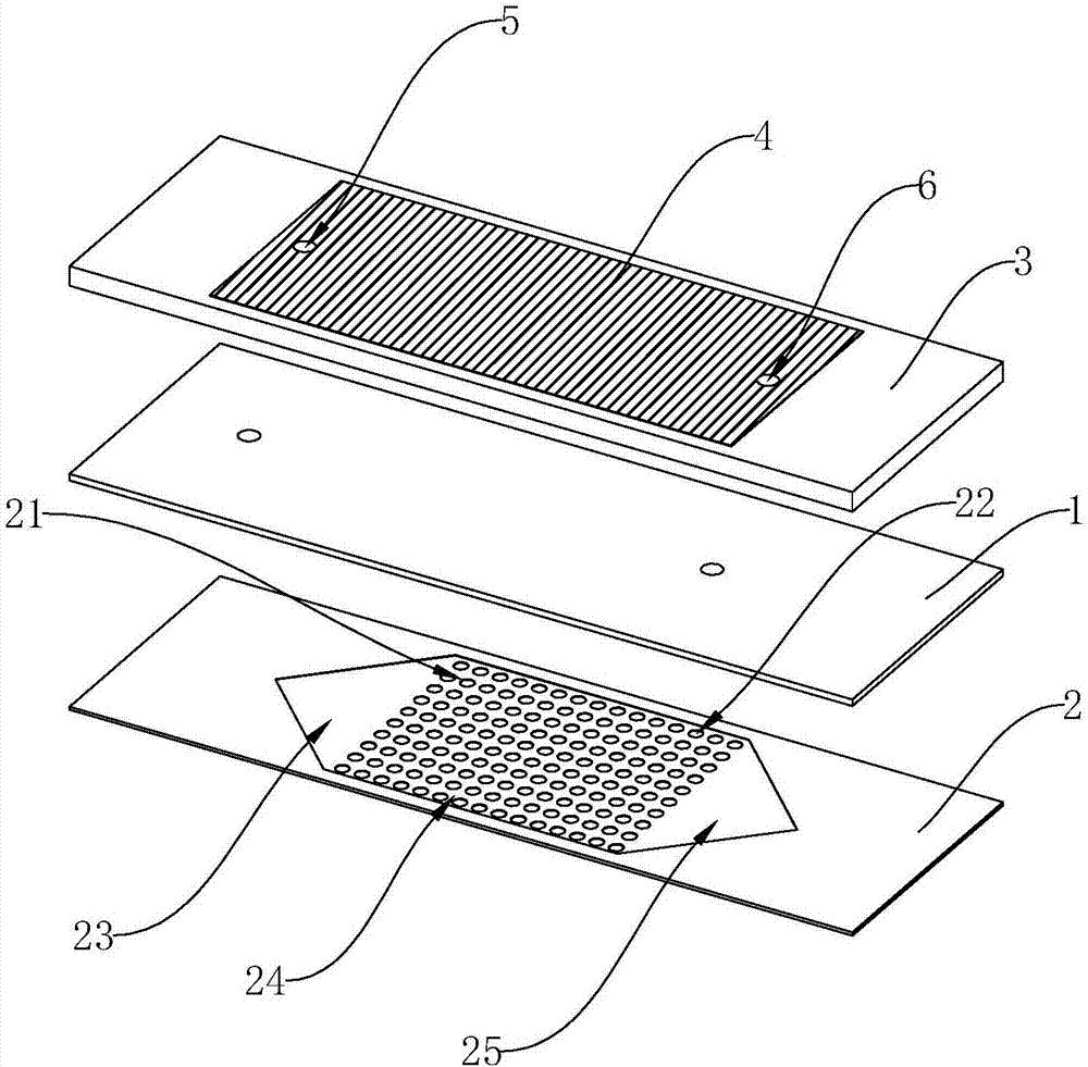
[0019] Embodiment 1: As shown in the figure, a biochip for screening, locating and detecting rare cells in blood includes a base layer 1 and a microfluidic channel layer 2, and the microfluidic channel layer 2 is closely arranged on the base layer 1. The upper surface of the microfluidic channel layer 2 is provided with a capture groove 21, and the bottom of the capture groove 21 is provided with a plurality of enrichment pits 22. The depth of the enrichment pits 22 is 15 to 50 microns. A signal amplification layer 3 is arranged close to the surface, and a nano-gold layer 4 is arranged on the upper surface of the signal amplification layer 3 corresponding to the capture groove 21, and an inlet channel 5 and a discharge channel 5 are provided through the nano-gold layer 4 and the signal amplification layer 3. The channel 6, the inlet channel 5 and the outlet channel 6 communicate with the catch tank 21, respectively.
Embodiment 2
[0020] Embodiment 2: As shown in the figure, a biochip for screening, positioning and detecting rare cells in blood includes a base layer 1 and a microfluidic channel layer 2, and the microfluidic channel layer 2 is arranged on the base layer 1 closely. The upper surface of the microfluidic channel layer 2 is provided with a capture groove 21, and the bottom of the capture groove 21 is provided with a plurality of enrichment pits 22. The depth of the enrichment pits 22 is 15 to 50 microns. A signal amplification layer 3 is arranged close to the surface, and a nano-gold layer 4 is arranged on the upper surface of the signal amplification layer 3 corresponding to the capture groove 21, and an inlet channel 5 and a discharge channel 5 are provided through the nano-gold layer 4 and the signal amplification layer 3. The channel 6, the inlet channel 5 and the outlet channel 6 communicate with the catch tank 21, respectively.
[0021] In this embodiment, the capture tank 21 is compos...
Embodiment 3
[0024] Embodiment 3: As shown in the figure, a biochip for screening, locating and detecting rare cells in blood includes a base layer 1 and a microfluidic channel layer 2, and the microfluidic channel layer 2 is closely arranged on the base layer 1. The upper surface of the microfluidic channel layer 2 is provided with a capture groove 21, and the bottom of the capture groove 21 is provided with a plurality of enrichment pits 22. The depth of the enrichment pits 22 is 15 to 50 microns. A signal amplification layer 3 is arranged close to the surface, and a nano-gold layer 4 is arranged on the upper surface of the signal amplification layer 3 corresponding to the capture groove 21, and an inlet channel 5 and a discharge channel 5 are provided through the nano-gold layer 4 and the signal amplification layer 3. The channel 6, the inlet channel 5 and the outlet channel 6 communicate with the catch tank 21, respectively.
[0025] In this embodiment, the capture tank 21 is composed ...
PUM
| Property | Measurement | Unit |
|---|---|---|
| depth | aaaaa | aaaaa |
| thickness | aaaaa | aaaaa |
| thickness | aaaaa | aaaaa |
Abstract
Description
Claims
Application Information
 - R&D
- Intellectual Property
- Life Sciences
- Materials
- Tech Scout
- Unparalleled Data Quality
- Higher Quality Content
- 60% Fewer Hallucinations
Browse by: Latest US Patents, China's latest patents, Technical Efficacy Thesaurus, Application Domain, Technology Topic, Popular Technical Reports.
© 2025 PatSnap. All rights reserved.Legal|Privacy policy|Modern Slavery Act Transparency Statement|Sitemap|About US| Contact US: help@patsnap.com


