Human amniotic epithelial stem cell serum-free medium and culture method thereof

A serum-free culture medium and serum-free culture technology, applied in cell dissociation methods, cell culture active agents, embryonic cells, etc., can solve the problems of non-immunogenicity and achieve strong clinical applicability, strong immunogenicity, The effect of overcoming the difficulty of obtaining materials

Active Publication Date: 2021-03-02
沈阳艾米奥生物工程技术研发中心有限公司
View PDF1 Cites 0 Cited by
  • Summary
  • Abstract
  • Description
  • Claims
  • Application Information

AI Technical Summary

Problems solved by technology

[0006] The present invention provides a serum-free culture medium for human amniotic membrane epithelial stem cells and a culture method thereof aiming at the problems existing in the above-mentioned prior art. Solve the problems of allogeneic, wide source, unrestricted by ethics and non-immunogenicity

Method used


Image

  • Human amniotic epithelial stem cell serum-free medium and culture method thereof
  • Human amniotic epithelial stem cell serum-free medium and culture method thereof
  • Human amniotic epithelial stem cell serum-free medium and culture method thereof

Examples


Embodiment 1

[0056] Such as figure 1 Shown, the schematic diagram of primary cultured hAESCs inverted microscope (×40). The primary amnion epithelial cells extracted from human amnion are uniform in shape and arranged like cobblestones.

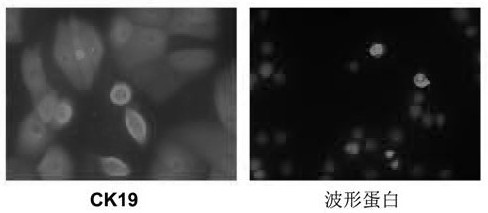
[0057] Such as figure 2 As shown, the cell immunofluorescence method was used to detect and identify the surface markers of the extracted hAESCs, and the expressions of the epithelial marker epithelial keratin CK19 (×100) and the mesenchymal cell marker vimentin (×100) in the P0 hAESCs extracted from the primary culture schematic diagram. The expression of CK19 was strongly positive and the expression of vimentin was weakly positive when hybridized with red fluorescent protein-labeled donkey anti-mouse fluorescent secondary antibody.

[0058] Such as image 3 As shown, the P1 passage hAESCs were cultured for 96 hours in serum-free medium (×40). Under the microscope, the cells were uniform in shape, and the cells were arranged like cobblestones.

[...

Embodiment 2

[0069] The serum-free medium of the human amniotic membrane epithelial stem cell bank of the present invention comprises:

[0070] Dulbecco's Modified Eagle's Medium / F12 (referred to as DMEM / F12) mixed by volume 1:1, 15.0~15.6g / L

[0071] Epidermal Growth Factor 0.005mg / L

[0072] Human transferrin 1.0mg / L

[0073] Human insulin 5.5mg / L

[0074] Sodium selenite 5.0×10 -3 mg / L

[0075] L-alanyl-L-glutamine dipeptide 300mg / L

[0076] L-alanine 10mg / L

[0077] L-Asparagine 4.9mg / L

[0078] L-Aspartic Acid 9.3mg / L

[0079] L-L-glutamic acid 10.7mg / L

[0080] Glycine 5.5mg / L

[0081] L-proline 7.5mg / L

[0082] L-serine 6.5mg / L

[0083] made into an aqueous solution.

Embodiment 3

[0085] The serum-free medium of the human amniotic membrane epithelial stem cell bank of the present invention comprises:

[0086] Dulbecco's Modified Eagle's Medium / F12 (referred to as DMEM / F12) mixed at a volume ratio of 1:1, 15.3g / L

[0087] Epidermal Growth Factor 0.01mg / L

[0088] Human transferrin 3.5mg / L

[0089] Human insulin 10mg / L

[0090] Sodium selenite 6.0×10 -3 mg / L

[0091] L-alanyl-L-glutamine dipeptide 400mg / L

[0092] L-alanine 17mg / L

[0093] L-Asparagine 7mg / L

[0094] L-Aspartic Acid 13mg / L

[0095] L-L-glutamic acid 14mg / L

[0096] Glycine 7.0mg / L

[0097] L-proline 10mg / L

[0098] L-serine 8mg / L

[0099] made into an aqueous solution.


PUM


Abstract

The invention relates to a serum-free medium of a human amniotic epithelial stem cell and a culture method thereof. The serum-free medium is formed by adding a human epidermal growth factor, human transferring, human insulin, sodium selenite, alanyl-L-glutamine dipeptide, alanine, asparaginate, aspartic acid, glutamic acid, glycine, proline and serine to a DMEM / F12 basic medium. The culture method comprises the steps of digesting 2.5g / L of trypsin for a human amniotic membrane, obtaining the human amniotic epithelial stem cell and filtering to prepare a single cell suspension; putting the human amniotic epithelial stem cell into a CO2 incubator of which the saturated humidity and the volume fraction are 5% at 37 DEG C through the serum-free medium under a serum-free condition and achieving growth and amplification of the amniotic epithelial stem cell through liquid exchange and subculture under the serum-free culture condition, the serum-free medium has the characteristics of stem cells, and is free of other animal sources, wide in source and free of ethical restriction.

Description

technical field [0001] The invention belongs to the field of biotechnology and relates to a serum-free culture medium for epithelial cells and a culture method thereof, in particular to a serum-free culture medium for human amniotic membrane epithelial cells and a culture method thereof. Background technique [0002] Human amnion is easy to obtain, does not cause ethical controversy, contains abundant amnion stem cells (amnion epithelial stem cells and amnion mesenchymal stem cells), and has low immunogenicity. It can become an important source of seed cells for clinical application of regenerative medicine. [0003] Human amniotic epithelial stem cells (hAESCs for short) are one of the stem cells isolated from amniotic membrane. hAESC is differentiated from the original two-germ layer embryo, which is developed from ectoderm cells on the 8th day after fertilization, earlier than the formation of three-germ layer embryos. This unique source of tissue embryology endows hAESC ...

Claims


Application Information

Patent Timeline
Patent Type & Authority Patents(China)
IPC IPC(8): C12N5/0735
CPCC12N5/0606C12N2500/05C12N2500/25C12N2500/32C12N2500/90C12N2501/11C12N2501/998C12N2509/00
Inventor 庞希宁施萍赵峰郎宏鑫
Owner 沈阳艾米奥生物工程技术研发中心有限公司
Why Eureka
Try Eureka