Eureka AIR delivers breakthrough ideas for toughest innovation challenges, trusted by R&D personnel around the world.

Three-way-decision-based liver tumor CT image classification method

A CT image, liver tumor technology, applied in the field of computer-aided medicine, can solve the problems of misclassification, inability to early warning of benign tumors, inability to obtain high classification accuracy, etc.

Inactive Publication Date: 2017-03-22
TONGJI UNIV
View PDF0 Cites 14 Cited by
  • Summary
  • Abstract
  • Description
  • Claims
  • Application Information

AI Technical Summary

Problems solved by technology

Its limitations are reflected in: for cases whose characteristics are not clear, it cannot achieve high classification accuracy, and it is easy to cause serious classification errors in judging difficult-to-distinguish malignant tumors as benign tumors; it cannot classify benign tumors with a high risk of cancer early warning

Method used


Image

  • Three-way-decision-based liver tumor CT image classification method
  • Three-way-decision-based liver tumor CT image classification method
  • Three-way-decision-based liver tumor CT image classification method

Examples


Embodiment 1

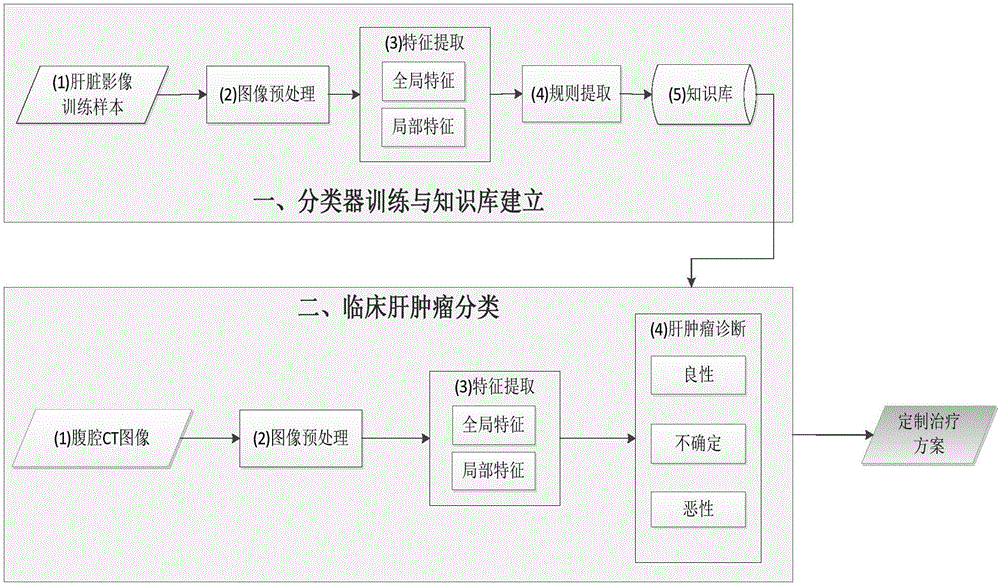
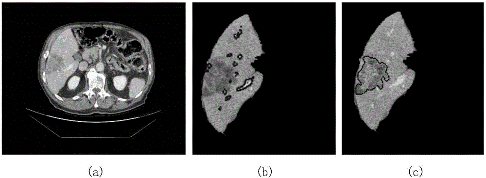
[0053] Refer to attached picture, in figure 1 A flow chart of the method of the present invention is provided in , and a group of embodiments are provided according to the flow chart of this diagram. This method first trains the classifier and labels 160 sets of liver CT image training samples, including 50 sets of liver cysts (benign), 50 sets of hepatic hemangiomas (benign) and 60 sets of hepatocellular carcinoma (malignant). The category attributes of the samples are divided into two categories: Class (benign / malignant). After image preprocessing and feature extraction, the sample forms a feature vector. On this basis, the attribute reduction method is used for rule extraction, and then a knowledge base is built to provide a basis for subsequent classification and diagnosis of new cases.

[0054] Image preprocessing includes extracting liver regions, liver vessels, and liver tumors. exist figure 2 In the example shown, the figure 2 (a) is a piece of original abdominal...


PUM


Abstract

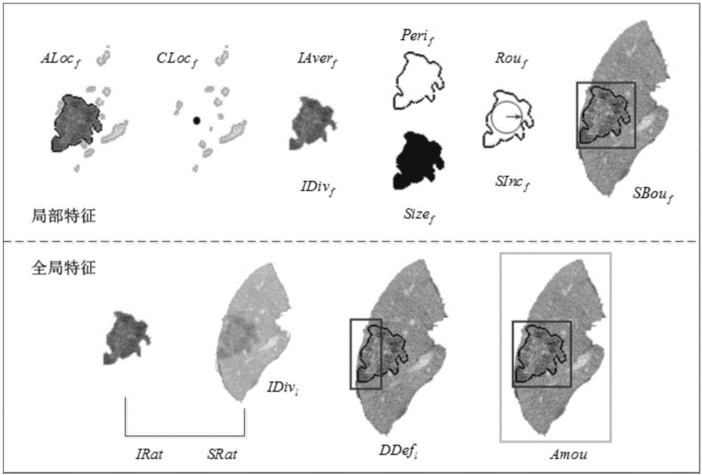
The invention discloses a three-way-decision-based liver tumor CT image classification method. A classifier is trained; image preprocessing and feature extraction are carried out on a marked liver CT image training sample to form a feature vector; rule extraction is carried out by using an attribute reduction method to form a knowledge base, thereby providing a basis for follow-up case classification diagnoses. T-be-classified cases are processed image pretreatment to extract liver regions, hepatic vessels, and liver tumors; 14 case features are calculated; the case feature values are inputted into a three-way-decision-based classifier; and then cases are classified into three types: a benign tumor, a malignant tumor, and an uncertain tumor. Therefore, a doctor can make different therapeutic regimens for different tumor types.

Description

technical field [0001] The invention relates to the field of computer aided medical treatment. Background technique [0002] Computer-aided classification of liver tumors (benign / malignant) plays a key role in the diagnosis and treatment of liver diseases. At present, the most common practice in hospitals is for doctors to classify the nature of tumors based on their experience and the characteristics of lesions in the two-dimensional image slice sequence. This diagnostic method is time-consuming, and classification accuracy largely relies on the subjective judgment of physicians. In recent years, the use of computer-based decision-making tools to aid clinical decision-making has been extensively studied and applied. However, the application of computer-aided liver tumor classification is less. Virmani Jitendra, Kumar Vinod, KalraNaveen, and Khandelwal Niranjan published an article titled Neural network based focal liver lesion diagnosis using image ultrasound in Computer...

Claims


Application Information

Patent Timeline
Patent Type & Authority Applications(China)
IPC IPC(8): G06T7/00G06K9/62
CPCG06T7/0012G06T2207/10081G06T2207/30096G06T2207/30056G06F18/24133G06F18/24
Inventor 陈宇飞岳晓冬龚晓亮
Owner TONGJI UNIV
Why Eureka
Eureka Blog
Learn More