


Quick Research
Generate reliable direction feasibility study reports for your R&D in just a few steps.

Technical Q&A
Discover and master advanced knowledge NOW. Basics, ideas, possibilities, all at once.

Find Solutions
As an expert in R&D theories, this can generate solutions to your technical problems instantly.

Evaluate Feasibility
Analyze your overall solution with one click, know your potential R&D risks in advance.

Monitor Landscape
Get weekly tech updates, stay abreast of the latest tech innovations and key insights.
Method for serum-free culture of cartilage cells and serum-free culture medium
A serum-free medium and chondrocyte technology, applied in the direction of cell culture active agent, bone/connective tissue cells, culture process, etc., can solve the problems of cell damage, chondrocytes that cannot be used for transplantation, and chondrocytes that cannot be well cultivated
- Summary
- Abstract
- Description
- Claims
- Application Information
AI Technical Summary
Problems solved by technology
Method used
Image

Examples
Embodiment 1
[0068] Digestion of Tissue - Culture Preparation -
[0069] Primary human chondrocytes were isolated individually from articular cartilage biopsies and the samples were subdivided. Then, immerse in a 0.1% to 0.3% collagenase solution, and perform enzyme digestion at 37° C. for 4 to 5 hours. The obtained cells were collected by centrifugation at 1,200 rpm for 5 minutes.
[0070] FGF 100 ng / mL, SAG 0.5 μM, HC 400 ng / mL, IGF 5 ng / mL and insulin 5 μg / mL were added to DMEM medium and used as a medium. Cultured cells previously obtained at 1000 cells / cm 2 seeding density. At 37°C, 5% CO 2 cultured in an environment.
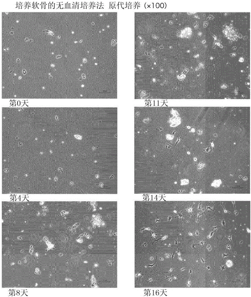
[0071] figure 1 It is a photograph in place of the drawing showing the cultured chondrocytes from the 0th day to the 16th day of culture in Example 1. It can be seen that the cartilage tissue increases slowly.
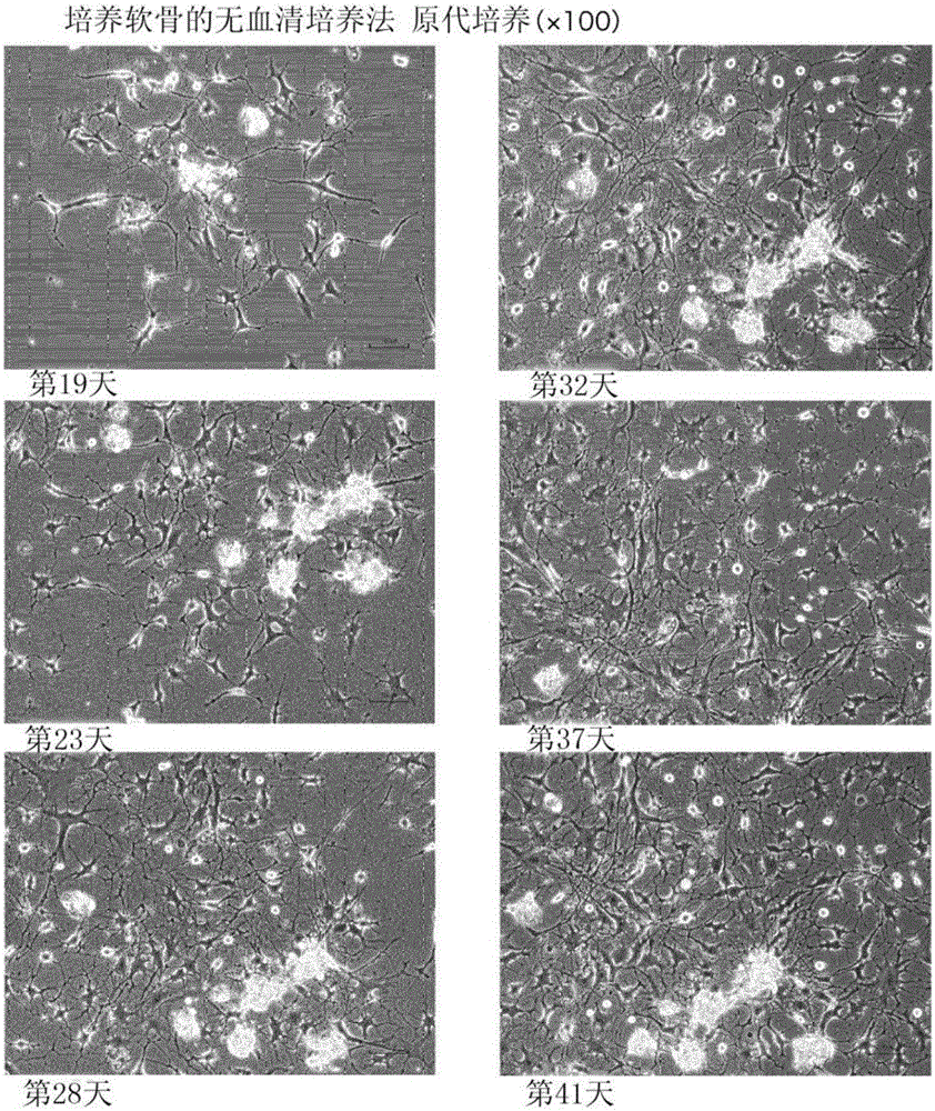
[0072] figure 2 It is a photograph in place of the drawing showing cultured chondrocytes from the 19th day to the 41st day of culture in Example 1. ...
Embodiment 2
[0087] FGF2 100 ng / mL, SAG 0.5 μM, HC 400 ng / mL, IGF 5 ng / mL, and insulin 5 μg / mL were added to DMEM medium to be used as a medium for subculture. The cultured cells in embodiment 1 were treated with 10000 cells / cm 2 seeding density. At 37°C, 5% CO 2 cultured in an environment.
[0088] Figure 4 It is a photograph in place of a drawing showing cultured chondrocytes from day 1 to day 13 in Example 2 (subculture). It was observed about 10 days from the start of the culture that stickiness was observed in the medium, and when the medium was removed with an aspirator, filaments were pulled out. From day 13, the viscosity of the medium increased.
[0089] Figure 5 It is a photograph in place of the drawing showing the cultured chondrocytes from the 18th day to the 28th day of culture in Example 2. On the 18th day of culture, 50 μg / mL of ascorbic acid was added to the medium. As a result of trial and error, it was found that the addition of ascorbic acid for such a period ...
Embodiment 3
[0098] In order to confirm that chondrocytes cultured in the serum-free medium of the present invention are cells suitable for transplantation, they were transplanted into nude mice to investigate whether cartilage was formed in vivo.
[0099] First, chondrocytes derived from human auricular cartilage were prepared according to the procedure described in Example 1, and cultured with the serum-free medium of the present invention. The cultured chondrocytes were impregnated in collagen cotton fabric, and transplanted together with the cotton fabric at two places on the back of nude mice. Transplantation surgery and management of nude mice were performed according to known methods. Six months after the transplantation, the back of the nude mouse was cut open, and it was confirmed whether the cells were fixed at the transplantation site and cartilage was formed.
[0100] Figure 8 This is a photograph showing the incision of the back of a nude mouse 6 months after transplantatio...
PUM
 Abstract
Description
Claims
Application Information
 - R&D Engineer
- R&D Manager
- IP Professional
- Industry Leading Data Capabilities
- Powerful AI technology
- Patent DNA Extraction
Browse by: Latest US Patents, China's latest patents, Technical Efficacy Thesaurus, Application Domain, Technology Topic, Popular Technical Reports.
© 2024 PatSnap. All rights reserved.Legal|Privacy policy|Modern Slavery Act Transparency Statement|Sitemap|About US| Contact US: help@patsnap.com