
Blood vessel recognition method and device

An identification method and technology of blood vessels, applied in medical science, laparoscopy, diagnostic signal processing, etc., can solve problems such as injury and damage to blood vessels, and achieve the effect of assisting surgery and diagnosis, assisting diagnosis, and reducing the risk of blood vessel damage.

Inactive Publication Date: 2016-09-28
THE FIRST AFFILIATED HOSPITAL OF XIAN JIAOTONG UNIV
View PDF10 Cites 6 Cited by
  • Summary
  • Abstract
  • Description
  • Claims
  • Application Information

AI Technical Summary

Problems solved by technology

Especially during surgery, if the position of the blood vessel is misjudged and the blood vessel is damaged, it will often lead to serious injury

Method used


Image

  • Blood vessel recognition method and device
  • Blood vessel recognition method and device
  • Blood vessel recognition method and device

Examples


Embodiment Construction

[0032] The present invention will be further described below in conjunction with the accompanying drawings.

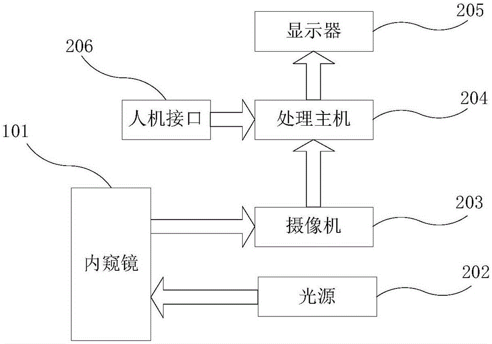
[0033] see figure 1 , a blood vessel identification method, comprising the following steps:

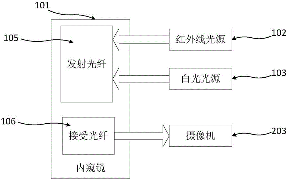
[0034] Step 1, the endoscope 101 is inserted into the abdominal cavity, illuminated by the white light source 103, and the infrared light source 102 of the endoscope 101 emits infrared rays to penetrate human tissues and absorb hemoglobin in the blood;

[0035] Step 2, shoot through the camera 203 in the endoscope 101, and the camera 203 sends the image information to the processing host 204 after filtering;

[0036] Step 3, the processing host 204 enhances the blood vessel image. The image enhancement adopts non-linear contrast transformation and image smoothing algorithm, especially when the blood vessel depth reaches about 5mm inside the tissue, an effective image enhancement method can provide convenience for subsequent processing;

[0037] Step 4, segment the enhanced b...


PUM


Abstract

The invention discloses a blood vessel identification method and equipment. By introducing a dual light source system (infrared light source + visible light source) and integrating the existing intelligent optimization algorithm optical system optical element adjustment method, the absorption ratio of infrared light to hemoglobin and other tissues is different. , combined with the optimization of computer algorithms, provides a blood vessel identification laparoscope, which can perform spectroscopic development with common endoscopes, achieve blood vessel identification, and is easy for doctors to use.

Description

technical field [0001] The invention belongs to the field of medical devices, and in particular relates to a blood vessel identification method and equipment. Background technique [0002] Laparoscope (English name: Laparoscopic) is a medical instrument with a miniature camera. Laparoscopic surgery is performed using laparoscope and related instruments: cold light source is used to provide illumination, and laparoscopic lens (diameter: 3-10mm) ) is inserted into the abdominal cavity, and digital camera technology is used to transmit the images captured by the laparoscopic lens to the post-stage signal processing system through the optical fiber, and display them on a dedicated monitor in real time. Then the doctor analyzes and judges the patient's condition through the images of different angles of the patient's organs displayed on the monitor screen, and uses special laparoscopic instruments to perform the operation. Laparoscopic surgery has less trauma, fewer complication...

Claims


Application Information

Patent Timeline
Patent Type & Authority Applications(China)
IPC IPC(8): A61B1/05A61B1/07A61B1/313A61B5/00
CPCA61B1/05A61B1/00186A61B1/0638A61B1/0661A61B1/07A61B1/3132A61B5/0086A61B5/489A61B5/6847A61B5/72
Inventor 王铮杨焜钟德星马磊仵正
Owner THE FIRST AFFILIATED HOSPITAL OF XIAN JIAOTONG UNIV
Learn More