
A kind of biological activity assay method of anti-PD-L1 monoclonal antibody

A technology of PD-L1 and monoclonal antibody, which is applied in the determination/inspection of microorganisms, biochemical equipment and methods, measuring devices, etc., can solve the problems of increasing patient mortality and enhancing tumor metastasis ability, and achieve good durability, Specific, precise and accurate results

Active Publication Date: 2018-08-07
兆科(广州)肿瘤药物有限公司
View PDF7 Cites 0 Cited by
  • Summary
  • Abstract
  • Description
  • Claims
  • Application Information

AI Technical Summary

Problems solved by technology

In tumor patients, high expression of PD-L1 can enhance tumor metastasis and lead to increased mortality

Method used


Image

  • A kind of biological activity assay method of anti-PD-L1 monoclonal antibody
  • A kind of biological activity assay method of anti-PD-L1 monoclonal antibody
  • A kind of biological activity assay method of anti-PD-L1 monoclonal antibody

Examples


Embodiment 1

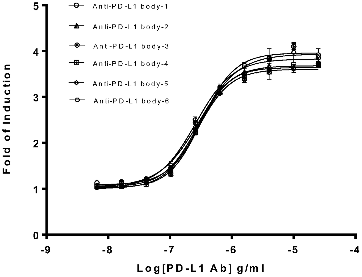
[0073] Example 1 Determination of the biological activity of 6 batches of different batches of anti-PD-L1 monoclonal antibodies

[0074] The 6 batches of different batches of anti-PD-L1 monoclonal antibodies are the anti-PD-L1 monoclonal antibodies produced by the company. The determination steps are as follows:

[0075] (1) Take out the cryopreserved PD1 cells and PD-L1 cells from the liquid nitrogen tank. After the cells are quickly thawed and recovered, they are transferred to a culture flask containing cell growth medium at 37°C, 5% CO 2 Cultivate in an incubator.

[0076] (2) Take PD-L1 cells in the logarithmic growth phase, digest and centrifuge, and dilute the cells to 4×10 5 Pcs / mL, then seeded in 96-well plate, seeding density is 4×10 per well 4 Pcs, at 37℃, 5% CO 2 Cultivate overnight in an incubator.

[0077] (3) Prepare 6 batches of different batches of anti-PD-L1 monoclonal antibody sample solutions, determine their concentration by BCA method, and use RPMI 1640 medium con...

Embodiment 2

[0084] Example 2 Using anti-PD-L1 monoclonal antibody to validate the method

[0085] Except for the special instructions in the following items, the experimental materials and methods are basically the same as the operations in Example 1.

[0086] 1 specificity

[0087] This experiment examines the specificity of target cells, effector cells and anti-PD-L1 monoclonal antibodies. Specifically:

[0088] 1.1 Target cell specificity: The effector cell is Jurkat / PD1, the antibody is anti-PD-L1 monoclonal antibody, and the target cells are CHO / PD-L1, CHO / PD-L1negative cell and Dilution buffer.

[0089] 1.2 Effector cell specificity: The target cell is CHO / PD-L1, the antibody is anti-PD-L1 monoclonal antibody, and the effector cells are Jurkat / PD1, Jurkat / PD1 negative cell and Dilution buffer.

[0090] 1.3 Specificity of anti-PD-L1 monoclonal antibody: the target cell is CHO / PD-L1, the effector cell is Jurkat / PD1, and the antibodies are: anti-PD-L1 monoclonal antibody, HER2 and Dilution buffe...


PUM

PropertyMeasurementUnit
correlation coefficientaaaaaaaaaa

Abstract

The invention relates to the field of activity detection of biological medicine, in particular to a biological activity determination method for an anti-PD-L1 monoclonal antibody. According to the method, a PD1 / PD-L1 interruption detection system is used, binding of PD1 and PD-L1 protein is interrupted when the anti-PD-L1 monoclonal antibody is bound with PD-L1 expressed by target cells, a transcription factor NFAT is activated through signal transduction, expression of luciferase is started, the expression quantity of luciferase is positively correlated to the biological activity of the anti-PD-L1 monoclonal antibody, the chemiluminescence value of the anti-PD-L1 monoclonal antibody is determined after a cell lysis solution and a luciferase substrate are added, a dose-effect curve is drawn, and accordingly the biological activity of the anti-PD-L1 monoclonal antibody is determined. The method is easy to implement, strong in specificity and high in durability and has great significance on quality control and clinical application of the anti-PD-L1 monoclonal antibody.

Description

Technical field [0001] The invention relates to the field of biopharmaceutical activity detection, in particular to a method for measuring the biological activity of an anti-PD-L1 monoclonal antibody. Background technique [0002] The PD-1 / PD-L1 signaling pathway is closely related to the immune escape of tumors and viruses, and is an important new target for immunotherapy of tumors and chronic infectious diseases. The use of monoclonal antibodies against PD-1 and PD-L1 to block the PD-1 / PD-L1 signaling pathway and restore the immune killing function of T cells can exert a good therapeutic effect and have a good application prospect. [0003] PD-1 is an immunosuppressive molecule that belongs to the CD28 family. It is a type I transmembrane protein composed of 268 amino acids and is located on the PDCD1 gene. Its structure mainly includes the extracellular immunoglobulin variable region (IgV) -Like domains, hydrophobic transmembrane regions and intracellular regions. The intracel...

Claims


Application Information

Patent Timeline
Patent Type & Authority Patents(China)
IPC IPC(8): C12Q1/66
CPCG01N33/577
Inventor 梁杰姚菲黄思伽戴向荣李小羿
Owner 兆科(广州)肿瘤药物有限公司
Learn More