Breast ultrasound scanning detection system

An ultrasonic scanning and detection system technology, which is applied in the field of breast ultrasonic scanning and detection systems, can solve the problems of inability to accurately locate breast examinations, and achieve the effects of small possibility, accurate positioning, and small breast deformation

Active Publication Date: 2018-03-30
CHISON MEDICAL TECH CO LTD
View PDF8 Cites 2 Cited by
  • Summary
  • Abstract
  • Description
  • Claims
  • Application Information

AI Technical Summary

Problems solved by technology

[0006] The purpose of the present invention is to overcome the problem in the prior art that the use of ultrasonic diagnostic equipment for mammary gland examination cannot be accurately positioned, and to provide a mammary gland ultrasonic detection system that can automatically rotate the probe for scanning and realize 360-degree omnidirectional stereoscopic imaging. Lesion localization is more accurate

Method used


Image

  • Breast ultrasound scanning detection system
  • Breast ultrasound scanning detection system
  • Breast ultrasound scanning detection system

Examples


Embodiment Construction

[0023] The present invention will be further described below in conjunction with drawings and embodiments.

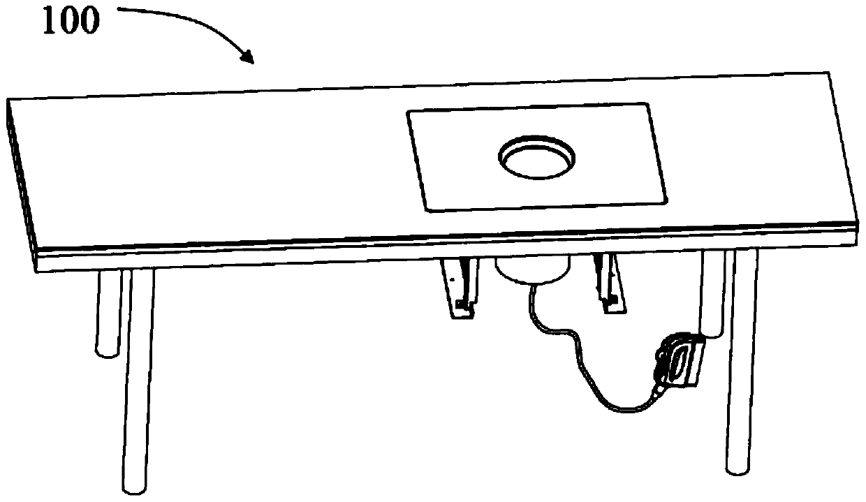
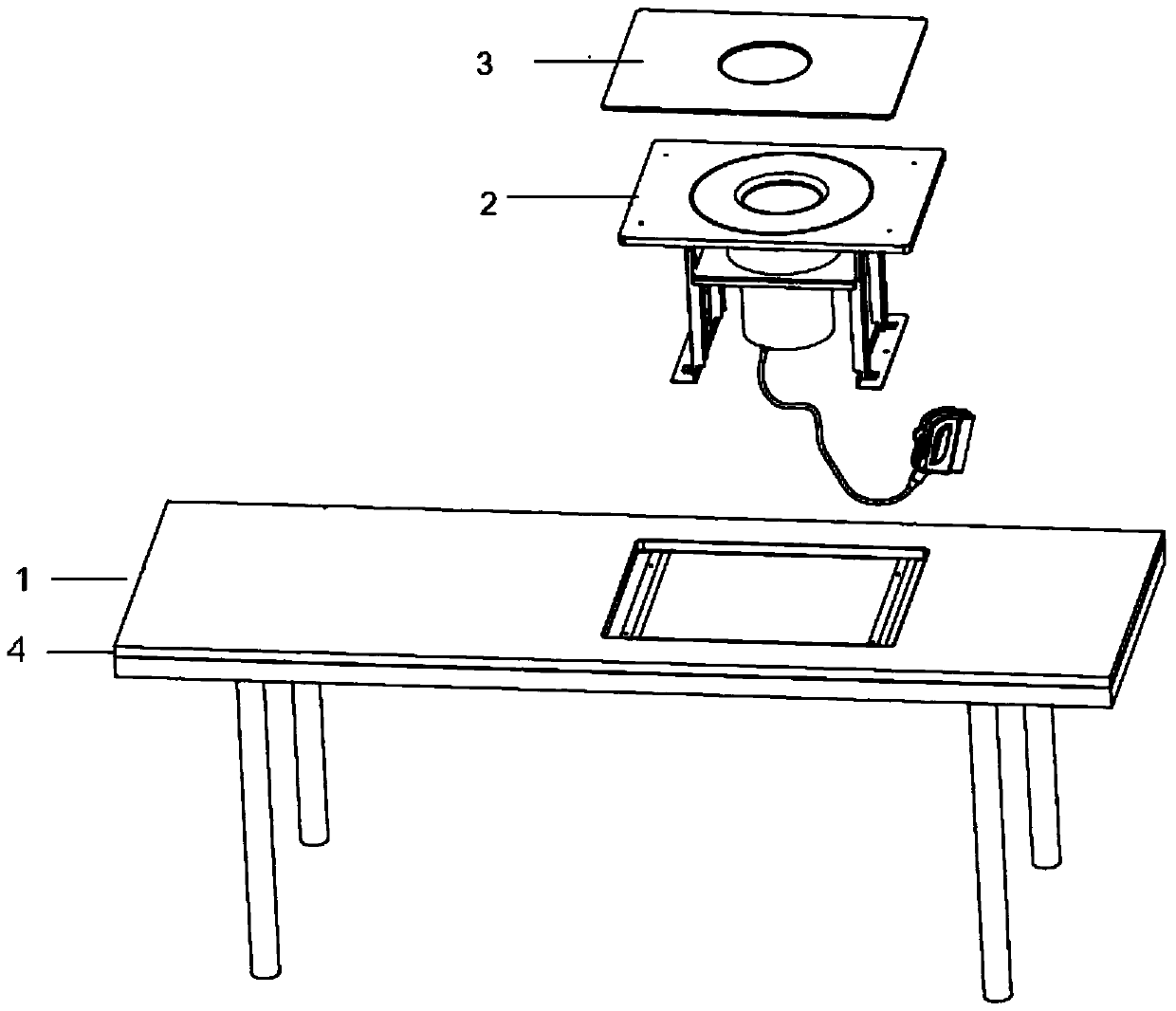
[0024] Such as figure 1 and 2 As shown, the present invention discloses a breast ultrasound scanning detection system 100, including an examination bed 1, an ultrasonic diagnostic instrument (not shown) and a scanning imaging device 2, the scanning imaging device 2 is installed on the examination bed 1, and is connected with Ultrasound connection. The scanning imaging device 2 is covered with a decorative cover plate 3 .

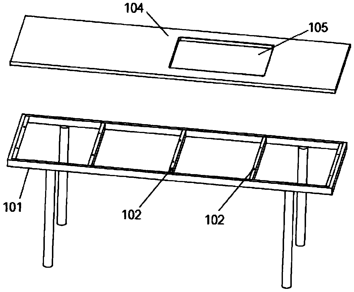
[0025] Such as image 3 As shown, the examination bed 1 includes a bed frame 101 , a beam 102 and a bed board 104 . The bed board 104 is provided with an inspection hole 105 for installing the scanning imaging device 2 . The bed board 104 is fixed on the bed frame 101, for example, by screws. The bed frame 101 and the beam 102 are assembled in a fixed connection, such as welding. In order to increase the stability of the examination bed 1, the ...


PUM


Abstract

The invention provides a mammary gland ultrasonic scanning detection system, which includes an examination bed, a scanning imaging device and an ultrasonic diagnostic instrument. A cover, a transducer, a driving device and a positioning device, the cup cover includes a cup body for holding interstitial fluid and accommodating the breast, a cup holder fixed below the cup body, and a cup holder for carrying the cup body and the cup holder. The cup holder and the cup holder are connected by bearings, and the cup holder is fixed to the examination table; the transducer is inserted into the cup body from the outside of the cup body; the upper end and the lower end of the driving device are respectively connected with the cup holder and the cup holder, and the driving device is equipped with Positioning device; when working, the cup body, the cup holder and the transducer form a rotating main body to make a rotary motion under the drive of the driving device. The present invention collects images completely through the fully automatic mode of the probe, which can more truly reflect the situation of the examiner and more accurately locate the lesion.

Description

technical field [0001] The invention relates to the technical field of ultrasonic imaging equipment, in particular to a mammary gland ultrasonic scanning detection system. Background technique [0002] Ultrasonic diagnostic equipment uses ultrasonic detection technology to understand the data and shape of human tissue structure through ultrasonic measurement. At present, the ultrasonic diagnostic instruments used in the industry generally include a main body and a probe. The probe is plugged into the main body and sends out ultrasonic waves through the transducer of the probe to scan and detect the target to be measured, and the obtained scanning data is fed back to the main body. Further processing is performed to form an intuitive image. [0003] At present, the application of ultrasonic diagnostic instruments in clinical diagnosis has been very popular, which has made great contributions to doctors' accurate understanding of patients' conditions and formulation of treatm...

Claims


Application Information

Patent Timeline
Patent Type & Authority Patents(China)
IPC IPC(8): A61B8/08A61B8/00
Inventor 宫明晶彭春林陆坚
Owner CHISON MEDICAL TECH CO LTD
Why Eureka
Try Eureka