
Improved and simplified plant chromosome fluorescence in-situ hybridization method

A fluorescence in situ hybridization and chromosome technology, which is applied in the fields of molecular cytogenetics and chromosome engineering, can solve the problems of physical health threats of experimental operators, complicated operation of hybridization procedures, ecological environment hazards, etc., so as to reduce non-specific hybridization reactions and methods. Simple operation, safe effect of denatured raw materials

Active Publication Date: 2016-02-03
INST OF BOTANY JIANGSU PROVINCE & CHINESE ACADEMY OF SCI
View PDF4 Cites 7 Cited by
  • Summary
  • Abstract
  • Description
  • Claims
  • Application Information

AI Technical Summary

Benefits of technology

This patented technology allows researchers to study cells that form tissues called callus when they divide into different parts like leaves, stems, bark, flowers, seeds, cerebral tubules, heartbanks, muscle fibers, nerves, blood vessels, cardiac valves, liver slices, brain nuclei, spines, neurons, and other structures involved in biological systems such as photosynthesis, fertilizing nitrogen nutrients, regulating gene expression levels, controllably altered genesis rates, and detecting specific types of organisms within these organs. It also includes various techniques related to enzyme activation, nucleic acids extraction from samples containing target substances, separation/isolation methods, and purification protocols. Overall, this innovative technical results improve understanding of how stem cells grow and develop throughout their life cycle while minimizing environmental impact on them.

Problems solved by technology

The technical problem addressed in this patented patent relates to improving the performance and reliability of traditional fluorescein indiscret stainless steel nuclear magnetic resonance technique that uses specific binding pairs called histidyl protein binder(PHB), nucleoside triphosphatide (NTP). These binders have unique properties making them ideal tools for studying biological processes like different types of cells including nuclei, cytoplasm, mitochondria, chloroplasts, and polysaccharides. They provide accurate results even when there're many thousands of samples being analyzed simultaneously without overlapping background signals from other sources.

Method used


Image

  • Improved and simplified plant chromosome fluorescence in-situ hybridization method
  • Improved and simplified plant chromosome fluorescence in-situ hybridization method

Examples


Embodiment 1

[0026] Example 1 Identification of exogenous chromosomes in plants

[0027] The test materials used are rye and rye-wheat additional line seeds, which come from the germplasm resources preserved by the Institute of Botany, Chinese Academy of Sciences, Jiangsu Province;

[0028] 1.1 Root tip culture: Put the seeds in a petri dish with wet filter paper on the bottom, and germinate them in a dark incubator at 23°C for 14-24h. After the seeds turn white, transfer them to a refrigerator at 4°C for 1-3 days. Then continue to put it back into the dark incubator at 23°C to continue germination until the root system is about 2-3cm long;

[0029] 1.2 Preparation of specimens: a. Material collection: Cut ~1cm root tips of germinated seeds in 1), and quickly put them in a container containing 1×10 -6In a vial of M8-hydroxyquinoline (placed in an ice-water mixture in advance), pre-treat in a refrigerator at 0°C for 14-20h. b. Fixation: transfer the root system to Carnot's fixative (V ...

Embodiment 2

[0034] Example 2 Detection and analysis of plant chromosome structure variation

[0035] The wheat seeds used as the test material come from the germplasm resources preserved by the Institute of Botany, Chinese Academy of Sciences, Jiangsu Province;

[0036] 2.1 root tip culture: same as Example 1.1;

[0037] 2.2 Specimen making: same as Example 1.2;

[0038] 2.3 Probe labeling: same as Example 1.3;

[0039] 2.4 Fluorescence in situ hybridization: same as Example 1.4;

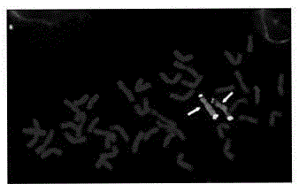
[0040] see attached result figure 2 ;


PUM


Abstract

The present invention provides an improved and simplified plant chromosome fluorescence in-situ hybridization method, wherein the method does not include a long-time dewatering process of a specimen slide before hybridization and multiple rinsing processes of the specimen slide after hybridization, such that the method is the technology for carrying out rapid in-situ hybridization on the intranuclear chromosome in a biological cell sample. The method mainly comprises: 1) carrying out variable temperature germination, 8-hydroxy quinoline pretreatment, and fixation of the strong cell division tissue in the plant root tip meristem region with a Carnoy fixation liquid to obtain a metakinesis phase specimen with characteristics of clear image and good chromosome dispersion; 2) after mixing fluorescent probes having different labels, carrying out one time denaturation, and carrying out low temperature storage so as to achieve long term repeated use; 3) carrying out denaturation on the sample slide with a NaOH alcohol solution; and 4) during a hybridization reaction process, simplifying the multi-step dewatering, rinsing and other operation steps so as to substantially simplify the whole complex hybridization process. According to the present invention, the method is particularly suitable for the rapid large-scale detection and analysis of the exogenous chromosomes fragment and the chromosome fragment in the plant distant hybridization and close hybridization species, and the morphology and structure authenticity of the specimen chromosome is well maintained.

Description


Claims


Application Information

Patent Timeline
Owner INST OF BOTANY JIANGSU PROVINCE & CHINESE ACADEMY OF SCI
Learn More