
Device and method for analyzing biomechanical property of cornea based on OCT three-dimensional imaging

A biomechanical and corneal technology, applied in the field of corneal biomechanical performance measurement, can solve the problems of undetectable corneal changes, unobtainable corneal changes, and undetectable corneal thickness changes.

Inactive Publication Date: 2016-01-13
深圳市亿领科技有限公司
View PDF8 Cites 11 Cited by
  • Summary
  • Abstract
  • Description
  • Claims
  • Application Information

AI Technical Summary

Problems solved by technology

However, there are the following deficiencies in the corneal expansion experiment: 1) only the displacement changes of the corneal apex can be obtained, but the overall changes of the cornea cannot be obtained; 2) the structural information such as the thickness and curvature of the cornea needs to be measured separately with other equipment; 3) The change of corneal thickness cannot be detected; 4) The local change of cornea cannot be detected

Method used


Image

  • Device and method for analyzing biomechanical property of cornea based on OCT three-dimensional imaging
  • Device and method for analyzing biomechanical property of cornea based on OCT three-dimensional imaging
  • Device and method for analyzing biomechanical property of cornea based on OCT three-dimensional imaging

Examples


Embodiment Construction

[0032] Below, the present invention will be described in detail with reference to the accompanying drawings.

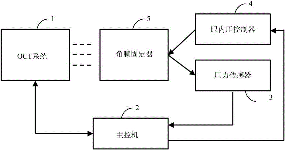
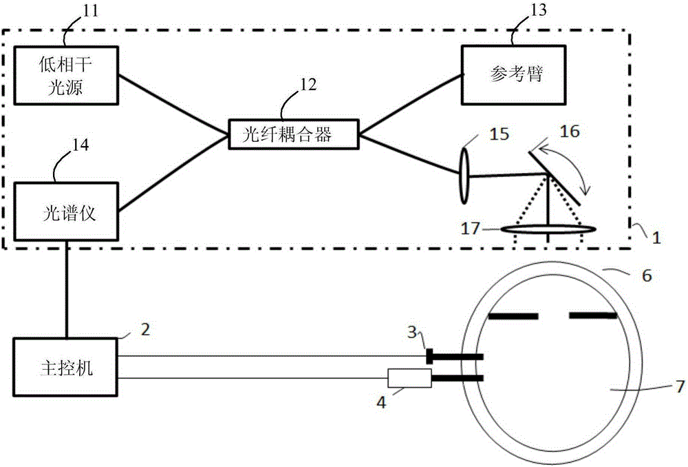
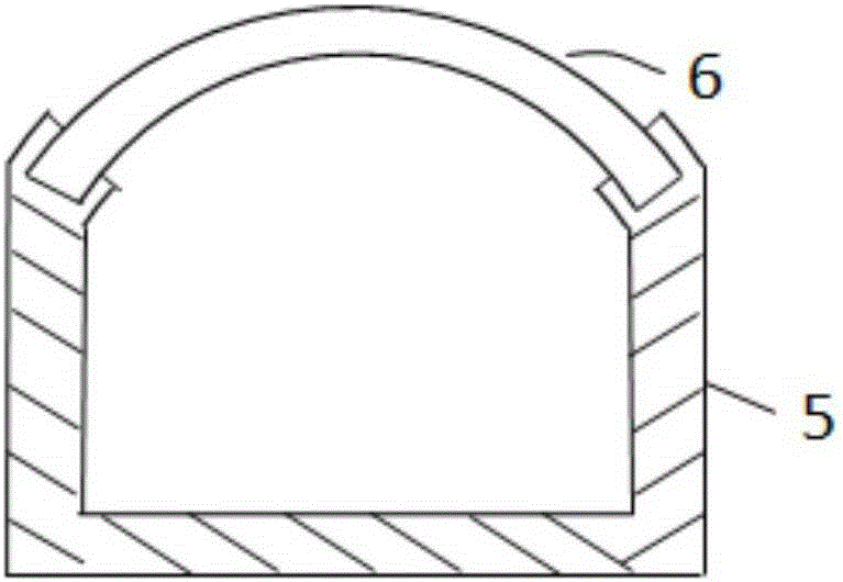
[0033] Such as Figure 1 to Figure 3 As shown, the present embodiment provides a device for analyzing corneal biomechanical properties based on OCT three-dimensional imaging, which device includes: OCT system 1, a master computer for controlling and measuring intraocular pressure and synchronously obtaining corneal OCT images 2. A corneal fixer 5 for fixing the isolated cornea, an intraocular pressure controller 4 connected with the corneal fixer 5 for controlling the pressure in the corneal fixer 5 and connected with the corneal fixer 5 for collecting the cornea 6. Pressure, i.e. pressure sensor force for intraocular pressure3.

[0034]Wherein, the OCT system 1 further includes: a low-coherence light source 11 , a fiber coupler 12 , a reference arm 13 , a spectrometer 14 , a fiber collimator 15 , a two-dimensional scanning galvanometer 16 and a converging lens 17 . ...


PUM


Abstract

The invention provides a device and method for analyzing the biomechanical property of cornea based on OCT three-dimensional imaging. The device comprises an OCT system, a master control machine, a cornea fixer, an intra-ocular pressure controller and a pressure sensor, wherein the OCT system comprises a low-coherence source, an optical fiber coupler, a reference arm, a spectrograph, an optical collimator, a two-dimensional scanning galvanometer and a syntaxis lens. According to the device and method, interference spectrum information is transmitted to the master control machine by virtue of the spectrograph, so that the cornea structure information is acquired after the master control machine is subjected to spectrum analysis, and parameters of the biomechanical property of cornea is acquired for disease diagnosis and researches.

Description

technical field [0001] The invention relates to the technical field of corneal biomechanical performance measurement technology, in particular to a device and method for analyzing corneal biomechanical performance based on OCT three-dimensional imaging. Background technique [0002] The cornea is the transparent tissue located at the front of the eyeball, providing protection for the eyeball and providing about 70% of the refractive function. The biomechanical properties of the cornea are related to the properties of the materials that make up the corneal tissue and the parameters of the corneal structure such as thickness and curvature. The biomechanical properties of the cornea are mainly reflected in the fact that the cornea can offset the intraocular pressure, maintain the shape of the cornea and protect the vision of the eyeball. At present, the biomechanical properties of the cornea are gradually being paid attention to in clinical medicine. For example, refractive sur...

Claims


Application Information

Patent Timeline
Patent Type & Authority Applications(China)
IPC IPC(8): A61B3/10A61B3/16A61B3/14
Inventor 王立科
Owner 深圳市亿领科技有限公司
Learn More