
X-ray and ultrasound combined breast examination device and fusion imaging method thereof

An X-ray and ultrasonic image technology, applied in the field of mammary gland inspection devices and its fusion imaging, can solve the problems of repeated equipment resource expenditure, incapable image processing, and low work efficiency, so as to shorten the time of mammary gland inspection, facilitate diagnosis, and improve work efficiency Effect

Inactive Publication Date: 2015-12-30
SHANTOU INST OF UITRASONIC INSTR CO LTD
View PDF4 Cites 9 Cited by
  • Summary
  • Abstract
  • Description
  • Claims
  • Application Information

AI Technical Summary

Problems solved by technology

[0002] When using ultrasound equipment to examine the target breast, it is necessary to compress the target breast to the inside of the patient's body and perform periodic vibrations to obtain automatic elastography, while when using X-ray equipment to examine the target breast, it needs to be rotated to obtain layered Therefore, at present, only X-ray equipment or ultrasound equipment can be used for mammary gland examination, and equipment resource expenditure is repeated, resulting in waste, long examination time, and low work efficiency.
[0003] In addition, ultrasound equipment inspection and X-ray equipment inspection generally require different body positions. The two inspection methods are carried out at different positions and deformations of the target breast. Therefore, the X-ray images obtained by X-ray equipment inspection, ultrasound equipment The breast ultrasound images obtained by the examination are two unrelated images, which cannot be further processed

Method used


Image

  • X-ray and ultrasound combined breast examination device and fusion imaging method thereof

Examples


Embodiment Construction

[0019] Further description will be given below in conjunction with the accompanying drawings and preferred embodiments of the present invention.

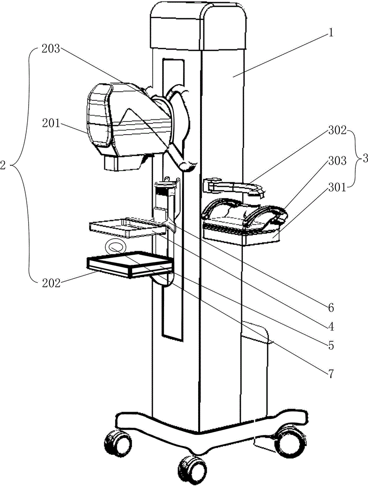
[0020] Such as figure 1 As shown, this mammary gland inspection device combining X-ray and ultrasound includes a frame 1, an X-ray inspection module 2, an ultrasonic inspection module 3, an upper compression plate 4, a lower compression plate 5, and a drive capable of driving the upper compression plate 4, The opening and closing mechanism 6 for the opening and closing movement of the lower pressing plate 5; the opening and closing mechanism 6 is installed on the frame 1, the upper pressing plate 4 and the lower pressing plate 5 are all installed on the power output end of the opening and closing mechanism 6, and the upper pressing plate 4 is above the lower compression plate 5; the X-ray inspection module 2 includes an X-ray emission source 201, an X-ray receiver 202 and a rotating mechanism 203 capable of driving the X-ray emissio...


PUM


Abstract

The invention relates to an X-ray and ultrasound combined breast examination device and a fusion imaging method thereof. The breast examination device comprises a rack, a rotatable X-ray examination module, an ultrasonic examination module capable of moving and reciprocating, an upper compression board, a lower compression board and an opening and closing mechanism which can drive the upper compression board and the lower compression board to open and close. Target breasts are fixed and compressed at the position of the breast examination device, the X-ray examination module and the ultrasonic examination module are adopted for examination, the positions and deformation of the target breasts are the same, ray image data and ultrasonic image data of corresponding points are overlaid to form a new breast image, the new breast image has the characteristics of a breast X-ray image and a breast ultrasonic image and facilitates diagnosis performed by a doctor, and the diagnosis result is accurate; the required image can be obtained by switching the X-ray examination module and the ultrasonic examination module, equipment resources are saved, the target breast image can be obtained more rapidly, and the working efficiency of the doctor is improved.

Description

technical field [0001] The invention relates to a mammary gland inspection device, in particular to a mammary gland inspection device combining X-rays and ultrasound and a fusion imaging method thereof. Background technique [0002] When using ultrasound equipment to examine the target breast, it is necessary to compress the target breast to the inside of the patient's body and perform periodic vibrations to obtain automatic elastography, while when using X-ray equipment to examine the target breast, it needs to be rotated to obtain layered Therefore, at present, only X-ray equipment or ultrasound equipment can be used for breast examination. The expenditure of equipment resources is repeated, resulting in waste, long examination time, and low work efficiency. [0003] In addition, ultrasound equipment inspection and X-ray equipment inspection generally require different body positions. The two inspection methods are carried out at different positions and deformations of the...

Claims


Application Information

Patent Timeline
IPC IPC(8): A61B8/00A61B6/00
Inventor 李德来周凌宏蔡泽杭鄞伟强赵榉云
Owner SHANTOU INST OF UITRASONIC INSTR CO LTD
Learn More