
A thoracoscopic surgery simulation training box

A surgical simulation and thoracoscopic technology, applied in teaching models, educational tools, instruments, etc., can solve the problems that trainees cannot be trained, and the structure and function are single

Inactive Publication Date: 2017-08-22
SOUTHERN MEDICAL UNIVERSITY
View PDF7 Cites 0 Cited by
  • Summary
  • Abstract
  • Description
  • Claims
  • Application Information

AI Technical Summary

Problems solved by technology

However, due to the single structure and function of this training box, trainees cannot receive the training closest to the real operating environment.

Method used


Image

  • A thoracoscopic surgery simulation training box
  • A thoracoscopic surgery simulation training box
  • A thoracoscopic surgery simulation training box

Examples


Embodiment Construction

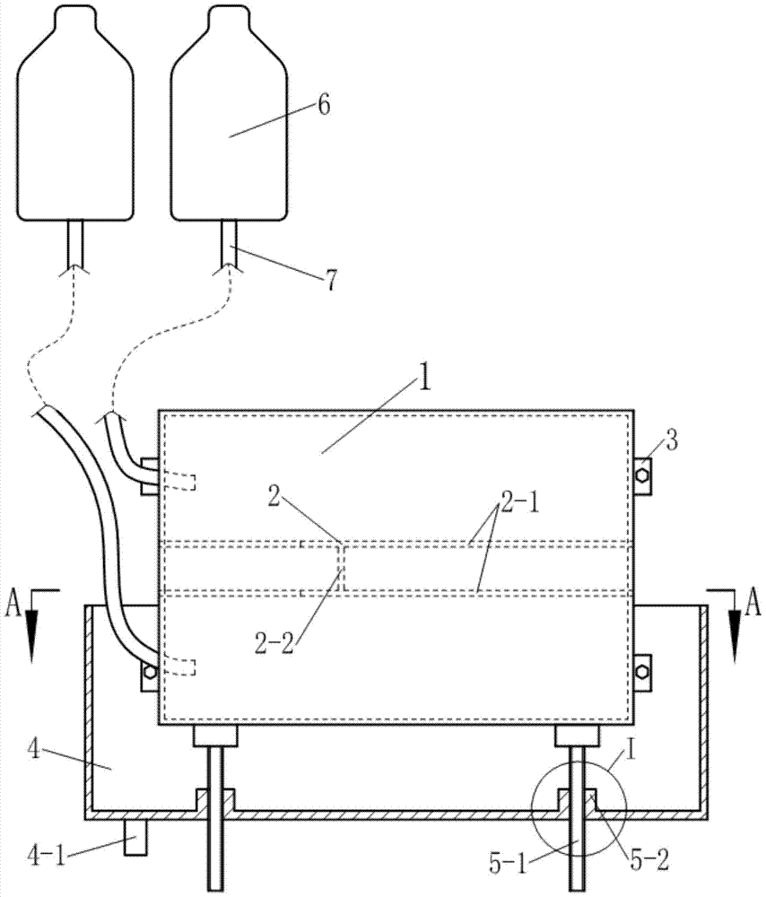
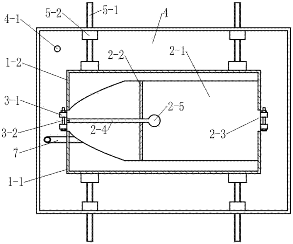
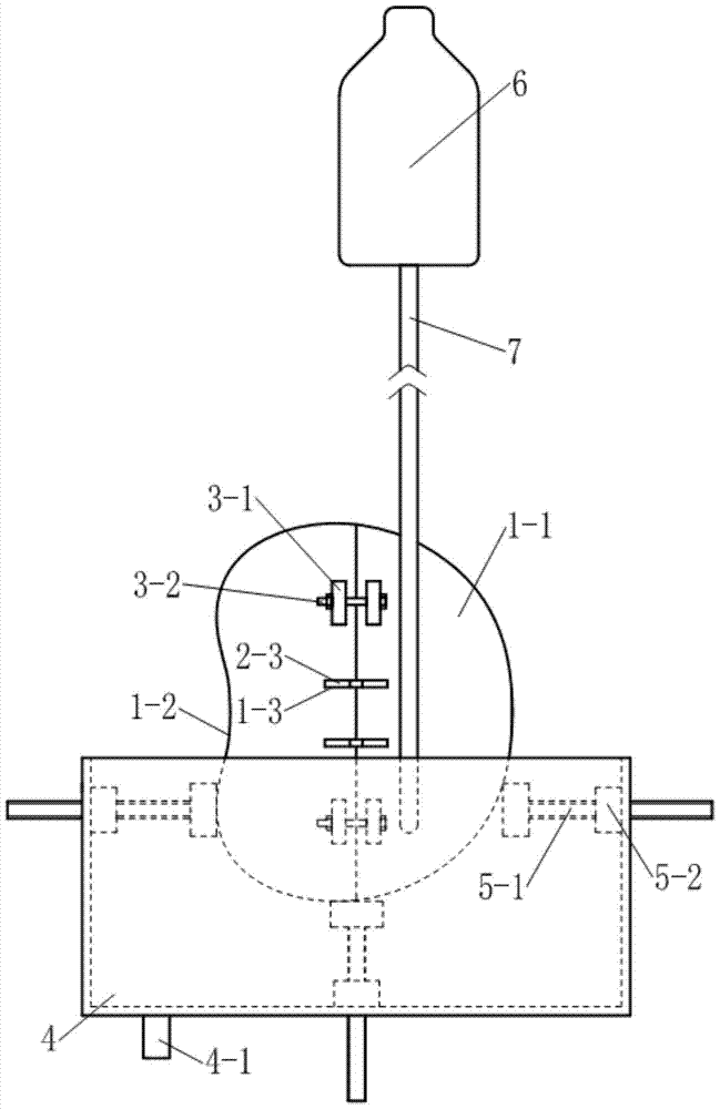
[0015] See Figure 1~6 In the example, the thoracoscopic surgery simulation training box is composed of a thoracic cavity prosthesis 1, a blood flow simulation device and a supporting device.

[0016] See Figure 1~4 The thoracic cavity prosthesis 1 is composed of a peripheral surface with a cross section similar to that of the thorax of the human body and partitions located at both ends of the peripheral surface. The thoracic cavity prosthesis 1 is divided into a front thoracic cavity prosthesis by the plane of the midaxillary line on both sides 1-1 and the posterior thoracic cavity prosthesis 1-2, the two are connected as a whole through the connecting device 3 provided at both ends of the thoracic cavity prosthesis 1. The connecting ear 3-1 on the prosthesis 1-2 and the bolt 3-2 threaded on the two connecting ears 3-1 are composed; the thoracic cavity prosthesis 1 is provided with a mediastinal pleural prosthesis 2, and the mediastinum The pleural prosthesis 2 is composed of ...


PUM


Abstract

The invention discloses a medical model, specifically a thoracoscopic operation simulation training box, the training box includes a chest prosthesis, and is characterized in that it also includes a blood flow simulation device and a supporting device; The cross-section is similar to the cross-section of the human thorax, and is divided into anterior and posterior thoracic prostheses by the plane of the midaxillary line on both sides; The mediastinal pleural plate on both sides and the connecting plate connecting the two; the blood flow simulation device is composed of two perfusion bottles and two perfusion catheters connecting the perfusion bottle and the thoracic prosthesis; the support device includes a A cuboid support box with an open top and support push rods on both sides and bottom of the support box. The bottom of the support box is provided with a perfusate recovery port. The push rod is composed of a push rod supported on the chest cavity prosthesis, and the push rod and the push rod seat are connected by threads.

Description

Technical field [0001] The invention relates to a medical model, which is used for simulating the operation environment and performing operation training of minimally invasive thoracic surgery. Background technique [0002] With the development of modern medical minimally invasive concepts, minimally invasive techniques have been more and more widely used in various surgical operations. Thoracoscopy is known as one of the major breakthroughs in thoracic surgery in the last century, and it is a representative operation of minimally invasive thoracic surgery. However, video-assisted thoracoscopic surgery is very different from conventional thoracotomy, and it is much more difficult. He is usually performed under 3-4 small chest wall incisions of about 1.5 cm. The doctor uses special surgical instruments to complete the operation by observing the endoscopic field of view displayed on the TV. However, after the images inside the patient's thoracic cavity are collected by laparoscop...

Claims


Application Information

Patent Timeline
Patent Type & Authority Patents(China)
IPC IPC(8): G09B23/28
Inventor 王昊飞孔祥雪聂兰英王雪莲李鉴轶王张林张辉见
Owner SOUTHERN MEDICAL UNIVERSITY
Learn More