
Image processing device, image processing system, and program

An image processing device and technology for capturing images, applied in image data processing, image data processing, image enhancement and other directions, can solve problems such as heavy workload, and achieve the effect of reducing the possibility of false detection

Inactive Publication Date: 2015-08-19
FUJIFILM CORP
View PDF8 Cites 7 Cited by
  • Summary
  • Abstract
  • Description
  • Claims
  • Application Information

AI Technical Summary

Problems solved by technology

Visual detection of NRBC is laborious due to the minimal amount of NRBC present in the blood of the uterus

Method used


Image

  • Image processing device, image processing system, and program
  • Image processing device, image processing system, and program
  • Image processing device, image processing system, and program

Examples


Embodiment Construction

[0039] Embodiments of the present invention will be described below with reference to the accompanying drawings.

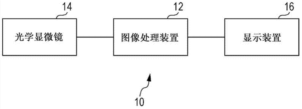
[0040] figure 1 is a configuration diagram showing an example of the image processing system 10 according to this embodiment. like figure 1 As shown, an image processing system 10 according to this embodiment includes an image processing device 12 , an optical microscope 14 and a display device 16 . The image processing device 12 is connected to the optical microscope 14 and the display device 16 by, for example, cables so as to exchange information with each other in a communicable manner.

[0041] For example, the optical microscope 14 according to this embodiment captures an image of a sample on colored glass set on a sample base by using a charge-coupled device (CCD) camera via an optical system such as an objective lens. In this example, the samples used were obtained by applying maternal blood to slides and subsequently applying a May-Giemsa stain thereto...


PUM


Abstract

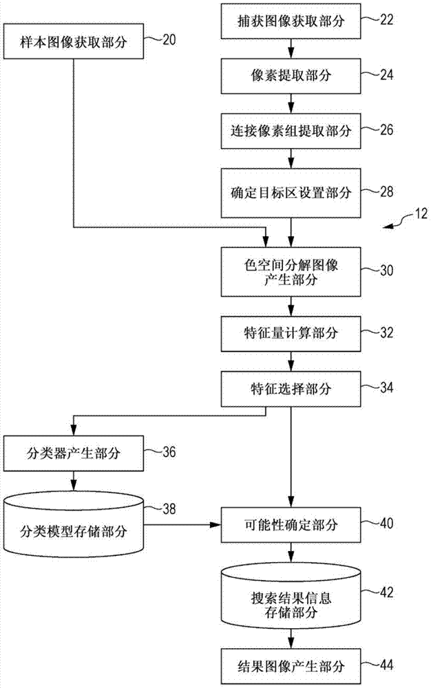
An objective of the present invention is to reduce, beyond a circumstance wherein this configuration is not present, the possibility of erroneous detection when searching for target cells by image processing from a captured image wherein target cells having nuclei are image captured. A captured image acquisition unit (22) acquires a captured image wherein a specimen including target cells having nuclei are image captured. A pixel extraction unit (24) extracts from pixels which are included in the captured image a pixel which is a nucleus candidate. A connected pixel group extraction unit (26) extracts a connected pixel group which is configured from pixels of a predetermined number of pixels or greater wherein mutually adjacent pixels are connected. On the basis of an image feature value which is identified on the basis of a brightness distribution for each of a plurality of mutually different color components within a region which encompasses at least a portion of the connected pixel group, and a condition of an image feature value which is mechanically learned on the basis of a brightness distribution for each of a plurality of mutually different color components within a sample image of the target cells, a possibility identification unit (40) identifies a value which denotes a possibility that the target cells are included in the regions.

Description

technical field [0001] The present invention relates to an image processing device, an image processing system, and a program. Background technique [0002] When diagnosing a fetus, the diagnosis is performed by detecting and utilizing an extremely small number of nucleated red blood cells (NRBCs, hereinafter referred to as "target cells") derived from the fetus and contained in the blood of the uterus. Since the number of NRBCs present in the blood of the uterus is extremely small, it is labor-intensive to detect NRBCs visually. As discussed in Patent Document 1, there is proposed a technique of mechanically detecting NRBCs by searching for cells conforming to conditions such as color, shape, positional relationship, and area ratio of NRBCs from a target image. [0003] Citation list [0004] patent documents [0005] PTL 1: Japanese Patent No.4346923 Contents of the invention [0006] technical problem [0007] An object of the present invention is to reduce the pos...

Claims


Application Information

Patent Timeline
Patent Type & Authority Applications(China)
IPC IPC(8): G06T1/00G06V10/50
CPCG06K9/4642G06K9/0014G06T2207/20076G06T2207/20081G06T7/11G02B21/244G06T2207/10056G06T2207/30024G06V20/695G06V10/50G06V20/698G06K2207/1012G06T7/0012G06T2207/10004G06T2207/10152G06T2207/30004G06T2207/30196
Inventor 尾崎良太
Owner FUJIFILM CORP
Learn More