
Kit and staining method for doubly staining mycobacterium tuberculosis and antigen of mycobacterium tuberculosis

A technology for Mycobacterium tuberculosis and a staining method is applied in the field of double staining kits for Mycobacterium tuberculosis and its antigens, which can solve the problems of low sensitivity, time-consuming and labor-intensive MTB, etc. The effect of clinical promotion value

Active Publication Date: 2015-05-06
BEIJING CHEST HOSPITAL CAPITAL MEDICAL UNIV
View PDF3 Cites 6 Cited by
  • Summary
  • Abstract
  • Description
  • Claims
  • Application Information

AI Technical Summary

Problems solved by technology

The most widely used MTB detection method is the Ziehl-Neelsen staining method, but there are disadvantages such as low sensitivity and time-consuming and labor-intensive search for MTB.

Method used


Image

  • Kit and staining method for doubly staining mycobacterium tuberculosis and antigen of mycobacterium tuberculosis
  • Kit and staining method for doubly staining mycobacterium tuberculosis and antigen of mycobacterium tuberculosis
  • Kit and staining method for doubly staining mycobacterium tuberculosis and antigen of mycobacterium tuberculosis

Examples


Embodiment 1

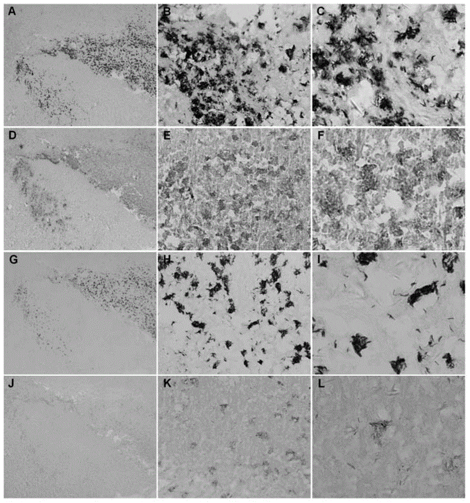
[0049] Embodiment 1, staining method of tuberculosis tissue specimen

[0050] 1. Reagent configuration:

[0051] 0.01mol / L, the sodium citrate buffer solution that pH value is 6.0: 2.41g sodium citrate (Na 3 C 6 h 5 o 7 2H 2 O) and 0.38g citric acid (C 6 h 8 o 7 ·H 2 O) Dissolve in 1 L of distilled water.

[0052] Concentration of 0.01M, pH value of 7.4 PBS formulation: 2.9g Na 2 HPO 4 12H 2 O, 0.3g NaH 2 PO 4 2H 2 O and 9gNaCl were dissolved in 1L of distilled water to adjust the pH value to 7.4.

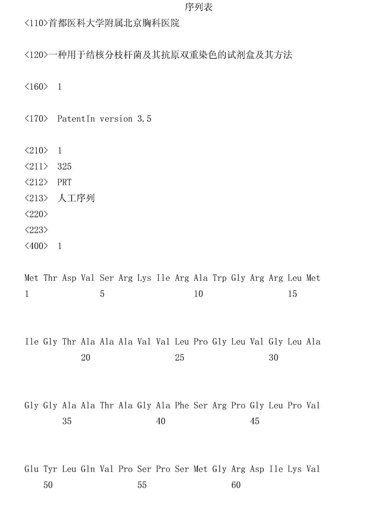
[0053] Polyclonal antibody against MTB antigen: New Zealand white rabbits were immunized with MTB antigen (amino acid sequence is sequence 1), rabbit serum was collected, and polyclonal antibody against MTB antigen was obtained by purification with proteinG.

[0054] DAB staining solution: first prepare 20×DAB dyeing mother solution: pour 0.1g of diaminobenzidine (3,3'-diaminobenzidine, DAB) into 10ml of distilled water to obtain a mixed solution, add 3-5 drops of 10M...

Embodiment 2

[0105] Embodiment 2, staining method of tuberculosis tissue specimen

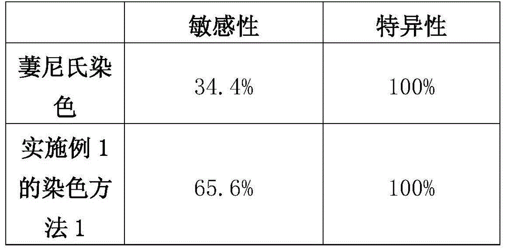
[0106] In order to explore the clinical application value of the kit prepared in Example 1, 212 clinically confirmed tuberculosis tissue samples (provided by Beijing Chest Hospital Affiliated to Capital Medical University, and informed by the patient) and 63 cases of control disease tissue samples (clinically confirmed) were collected. The non-tuberculosis tissue samples were provided by Beijing Chest Hospital Affiliated to Capital Medical University, and the patients were informed).

[0107] Above-mentioned sample is tested according to dyeing method 1 shown in embodiment 1;

[0108] At the same time, traditional lindensl staining was used as the control.

[0109] Traditional method of linseed and Nissl staining:

[0110] 1) Prepare 4 μm thick paraffin tissue sections, routine xylene dewaxing, gradient alcohol hydration.

[0111] 2) Add phenol fuchsin staining solution dropwise, cover the tissue to be t...


PUM


Abstract

The invention discloses a kit and a staining method for staining mycobacterium tuberculosis and the expression protein of the mycobacterium tuberculosis. The method for staining the mycobacterium tuberculosis and the expression protein of the mycobacterium tuberculosis comprises the following steps: (1) carrying out primary antibody incubation and secondary antibody incubation to a to-be-tested sample containing mycobacterium tuberculosis to obtain an antibody binding product; and (2) carrying out DAB (diaminobenzidine) staining solution staining, carbolfuchsin staining solution staining and hematoxylin staining solution staining to the antibody binding product sequentially, so as to obtain stained mycobacterium tuberculosis and the stained expression protein. Experimental results show that a novel kit for the tuberculotic pathology diagnosis is developed, and the kit can detect the mycobacterium tuberculosis and the expression protein in one tissue specimen section simultaneously, so as to improve the tuberculotic pathological diagnosis sensibility. The kit can be used conveniently and simply, is suitable for the conventional detection for the pathology department and has good clinical promotion value.

Description

technical field [0001] The invention relates to the field of biotechnology, in particular to a kit for double staining of mycobacterium tuberculosis and its antigen and a method thereof. Background technique [0002] Tuberculosis is an infectious disease that seriously threatens human health. One-third of the world's population is infected with Mycobacterium tuberculosis (MTB), and 2 million people die from tuberculosis every year. The death rate is only the lowest among infectious diseases. Ranked second to AIDS. The current gold standard for TB diagnosis is sputum bacteriology. However, the positive rate of sputum smears is low, and it takes one month for sputum culture to produce results. Therefore, the most commonly used sputum bacteriological detection methods cannot meet the needs of clinical diagnosis. In addition, in recent years, new technologies such as ELISPOT based on immunology and PCR based on nucleic acid detection have also been applied to the diagnosis of ...

Claims


Application Information

Patent Timeline
IPC IPC(8): G01N1/30G01N33/569
Inventor 车南颖张海青
Owner BEIJING CHEST HOSPITAL CAPITAL MEDICAL UNIV
Learn More