
Assessment of patency using photoplethysmography on endoscopic images

A technology of endoscopy and images, applied in the direction of endoscopy, application, diagnostic recording/measurement, etc., can solve the problems that surgeons cannot obtain, flow blockage, etc.

Active Publication Date: 2017-09-08
KONINKLJIJKE PHILIPS NV
View PDF0 Cites 0 Cited by
  • Summary
  • Abstract
  • Description
  • Claims
  • Application Information

AI Technical Summary

Problems solved by technology

However, successful revascularization of an artery may not necessarily result in successful perfusion of the myocardium since flow in smaller vessels (which are not visible on conventional imaging) may also be able to be occluded
Additionally, these imaging modalities are often not available to surgeons

Method used


Image

  • Assessment of patency using photoplethysmography on endoscopic images
  • Assessment of patency using photoplethysmography on endoscopic images
  • Assessment of patency using photoplethysmography on endoscopic images

Examples


Embodiment Construction

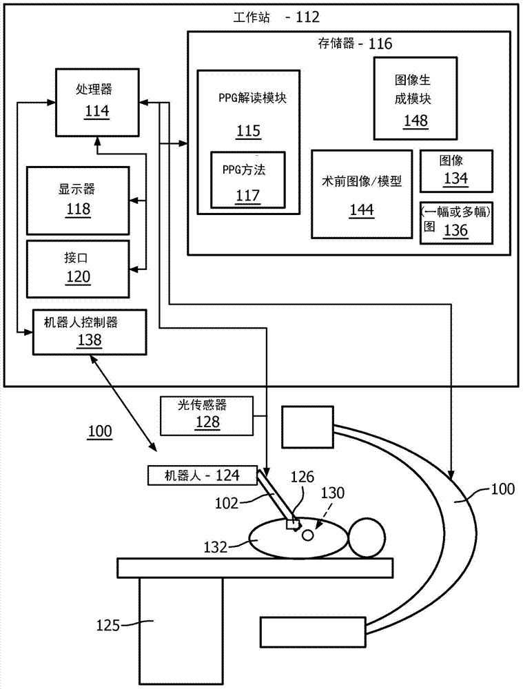
[0019] In accordance with the principles of the present invention, systems and methods are provided for determining fluid flow in a region of interest using light emitted or reflected from tissue. In one embodiment, photoplethysmography (PPG) is used to assess blood flow in tissue. PPG uses light reflection or transmission to detect cardiovascular pulse waves passing through the body. PPG is based on the principle that blood absorbs more light than surrounding tissue, so changes in blood volume affect transmission or reflection accordingly. The PPG signal can be used to detect respiration rate and heart rate using only a CCD camera and ambient light illumination. The systems and methods described herein can extract, for example, green and blue pixel intensities from a region of interest on a CCD camera-based image, and then measure their changes over time. Other information can also be extracted and monitored. A higher amplitude signal corresponds to a higher reflectivity a...


PUM


Abstract

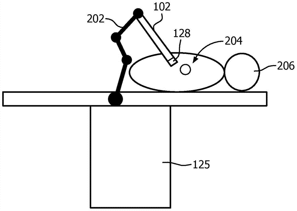
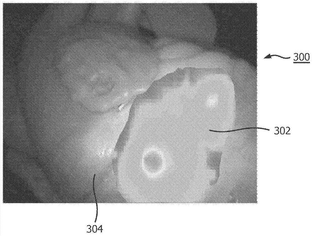
A system for evaluating patency includes a light sensor (128) positionable relative to a blood vessel to receive light from the blood vessel and convert the light into an image signal. A photo-plethysmography (PPG) interpretation module (115) is configured to receive the image signal and output pixel values in an image representing PPG information. An image generation module (148) is coupled to the PPG interpretation module to receive the pixel values and generate a PPG map to be output to a display for analysis.

Description

technical field [0001] The present disclosure relates to medical devices and procedures, and more particularly to monitoring fluid flow in tissue using photoplethysmographic (PPG) information. Background technique [0002] During heart bypass surgery, surgeons use one or more blood vessels from the body to bypass atherosclerotic patients with narrowed coronary arteries. The intent of this procedure is to increase blood flow in the coronary arteries, which supply the heart's myocardial tissue. During minimally invasive (MI) heart bypass surgery, an elongated instrument is inserted into a small incision in the patient's chest, and an endoscope provides visualization. In robotic-guided MI cardiac bypass surgery, one or more of the instruments (or endoscopes) are controlled by a robotic device. Heart bypass surgery can be performed while the patient is on cardiopulmonary bypass and the heart is in cardiac arrest, or while the heart is beating. [0003] X-ray angiography or ul...

Claims


Application Information

Patent Timeline
Patent Type & Authority Patents(China)
IPC IPC(8): A61B5/00A61B1/00A61B5/0295
CPCA61B34/30A61B1/0005A61B5/0084A61B5/0261A61B5/0295A61B5/061A61B5/1459A61B5/4851A61B5/7285A61B2034/301
Inventor H·埃尔哈瓦林A·波波维奇W·韦尔克鲁杰塞
Owner KONINKLJIJKE PHILIPS NV
Learn More