Method for screening a whitening substance using mitochondrial dynamics, and kit using the method
A screening method, mitochondrial technology, applied in the direction of biochemical equipment and methods, scientific instruments, microbial determination/examination, etc., can solve the problem of not showing the inhibitory effect of human skin hyperpigmentation, not easy to skin tissue, a lot of cost and effort And other issues
- Summary
- Abstract
- Description
- Claims
- Application Information
AI Technical Summary
Problems solved by technology
Method used
Image

Examples
Embodiment 1
[0051] Example 1: Induction of Mitochondrial Fusion Using Drugs
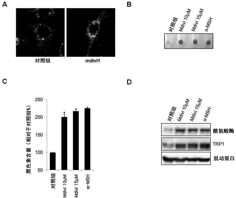
[0052] mouse melanoma B16F1 cells (purchased from ATCC), 1.6×10 4 Cell / ml concentration, placed in 12-well plate. After 24 hours, they were treated with 10 μM and 15 μM Mdivi-1 (Enzo Life Sciences) and 200 nM α-MSH (positive control group for melanin induction). After the fusion was induced by treating Mdivi1, observed with confocal laser microscope, it can be observed as figure 2 A shows the fusion of mitochondria. Cells were harvested after 48 hours and lysed with 1N NaOH at 100°C for 30 minutes. Afterwards, the cells were collected, and the total amount of melanin was observed with the naked eye, which was compared with figure 2 B shows the same. Measure the absorbance (SpectraMax190 microplate reader, Molecular Instruments Corporation of America, the U.S.) at 470nm afterwards. figure 2 The total amount of melanin measured by quantifying the results of B is shown in the graph of Fig. 2C. Also, b...
Embodiment 2
[0053] Example 2: Induction of Mitochondrial Fusion Using DRP1 siRNA
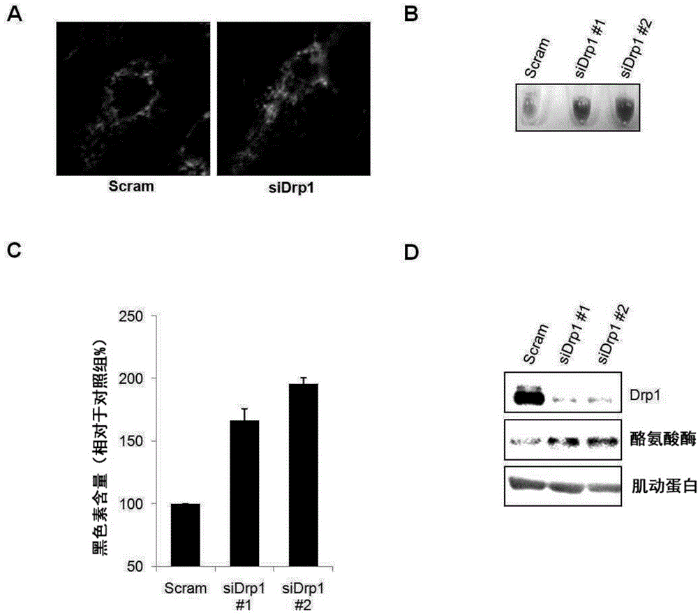
[0054] Mouse melanoma B16F1 cells, 1.6×10 4 Cells / ml concentration, placed on a 12-well plate. 24 hours later, DRP1 siRNA #1100 pmol was transfected using liposome reagent (Invitrogen). After treatment with DRP1siRNA to induce fusion, observe with a laser confocal microscope (LSM510, Carl Zeiss (Carl Zeiss), USA), and it can be observed that image 3 A shows the fusion of mitochondria. Cells were harvested after 72 hours and lysed with 1N NaOH at 100°C for 30 minutes. Afterwards, the cells were collected, and the total amount of melanin was observed with the naked eye, which was compared with image 3 B shows the same. Then the absorbance was measured at 470nm, and the figure 2 The results of B are quantified to measure the total amount of melanin, using image 3 The graph of C shows. Also, by Western blotting image 3 B shows the amount of the melanogenesis-regulating protein inside the cell afte...
Embodiment 3
[0057] Example 3: Induction of Mitochondrial Fission Using Drugs
[0058] Mouse melanoma B16F1 cells, 1.6×10 4 Cells / ml concentration, placed on a 12-well plate. After 24 hours, the cells were treated with 2.5 μM and 5 μM CCCP (Sigma) and 200 nM α-MSH. At this time, CCCP was treated under the condition that treatment of α-MSH induces melanin biosynthesis. In addition, after treatment with CCCP alone, after inducing division, observe with a laser confocal microscope (LSM510, Carl Zeiss, USA), it can be observed as Figure 4 A shows the division of mitochondria. After 48 hours, the cells were harvested and lysed at 100° C. for 30 minutes with 1N NaOH. Afterwards, the cells were collected, and the total amount of melanin was observed with the naked eye, which was compared with Figure 4 B shows the same. Measure the absorbance (SpectraMax190 microplate reader, Molecular Instruments Corporation of America, the U.S.) at 470nm afterwards. Figure 4 The results of B are quanti...
PUM
 Abstract
Description
Claims
Application Information
 - Generate Ideas
- Intellectual Property
- Life Sciences
- Materials
- Tech Scout
- Unparalleled Data Quality
- Higher Quality Content
- 60% Fewer Hallucinations
Browse by: Latest US Patents, China's latest patents, Technical Efficacy Thesaurus, Application Domain, Technology Topic, Popular Technical Reports.
© 2025 PatSnap. All rights reserved.Legal|Privacy policy|Modern Slavery Act Transparency Statement|Sitemap|About US| Contact US: help@patsnap.com