
Preparation method for hydroxyapatite nanotube and application to bone restoration

A hydroxyapatite and nanotube technology, applied in medical science, prosthesis, etc., can solve the problems of limited application of bone transplantation, infection in the bone extraction area, limited amount of bone donation, etc., achieving low cost, enhanced antibacterial ability, operation handy effect

Inactive Publication Date: 2015-01-21
郑欣
View PDF7 Cites 7 Cited by
  • Summary
  • Abstract
  • Description
  • Claims
  • Application Information

AI Technical Summary

Benefits of technology

In this patented method, we developed an improved way to create stable biocompatible scaffolds that are suitable for medical applications like dental implants. These scents consist mainly of calcium phosphate particles mixed together into a gel material made from polymers called Poly vinyl Alcohol). They were found effective at preventing inflammation caused by plaque buildup on teeth due to their high affinity towards acid proteins present naturally occurring substances commonly associated with periodontitis. Additionally, these scents could release other drugs through interactions between them and promote cellular growth within natural environments where they reside during metabolism. Overall, these technical features make these scorbents useful tools for medicine research purposes.

Problems solved by technology

This patented technical problem addressed in this patents relates to developing an effective way to treat damaged areas due to severe trauma during orthopaedics procedures without causing any harm from remaining foreign bodies like catheters. Current methods involve removing all healthy parts of the patient's own joint before injecting immunosuppressants which may be associated with increased risks of secondary injury after reconstructing soft tissue repairs. Therefore, current therapies require extensive surgeries and donors, making them challenging targets for future regenerative medicine approaches aiming towards more natural sources of replacement materials.

Method used


Image

  • Preparation method for hydroxyapatite nanotube and application to bone restoration
  • Preparation method for hydroxyapatite nanotube and application to bone restoration
  • Preparation method for hydroxyapatite nanotube and application to bone restoration

Examples


Embodiment 1

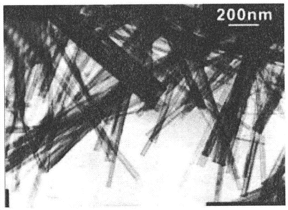
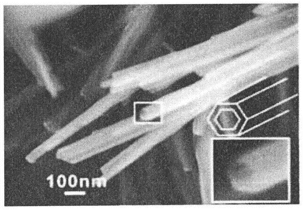
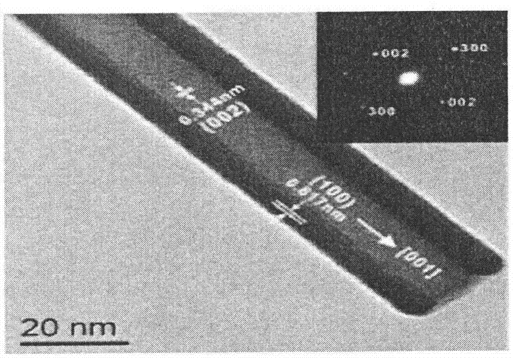
[0048]Take 100 mg of composite material sample and immerse in 4 mL of phosphate buffer (pH=7.4), and then place it in a 37°C incubator. Take out the sample 24 hours after soaking, centrifuge at 8000r / min for 15 minutes, absorb the leaching solution, wash the tube wall once with PBS solution, replace the PBS solution, and continue soaking. The concentration of BMP-7 in the leachate sample was detected by spectrophotometer, and all experiments were carried out under aseptic operation. Finally, the BMP-7 content released from the composite material was calculated and fitted with a drug release model. Electron micrographs of hydroxyapatite nanotubes loaded with gentamicin before and after release. ( Figure 8 , 9 , 10, 11)

Embodiment 2

[0050] Fifteen male New Zealand rabbits aged 6 months were selected, injected intramuscularly with ketamine 50mg / kg, anesthetized with 3% sodium pentobarbital 20mg / kg ear margin veins, after successful anesthesia, prepared the skin on both forearms, and fixed them on the rabbit board in the supine position , Disinfect the operating area, and spread sterile drapes. Cut the skin, subcutaneous tissue and fascia at the upper radial side of the forearm, separate the muscles to expose the periosteum of the radius, remove the periosteum of the radius with a surgical blade, and then use an orthopedic electric drill loaded with a grinding wheel with a diameter of 1 cm to remove the periosteum of the radius. Radius resection was performed to remove the periosteum, and the ulnar periosteum of the resected radius was carefully separated to create a bone defect with a defect length of 1.5 cm. Pay attention during the operation that the two sections of the radial bone defect are parallel an...


PUM

PropertyMeasurementUnit
Diameteraaaaaaaaaa

Abstract

The invention aims at providing a hydroxyapatite nanotube which possesses drug loading capacity and has the aperture of about 100-200 nm. The hydroxyapatite nanotube is prepared by utilizing cheap easily-available nanometer hydroxyapatite (HA) powder, polyvinyl alcohol (PVA) and polyvinylpyrrolidone (PVP) and employing a three-dimension printing technology. The hydroxyapatite nanotube with bionic functions is designed by utilizing the three-dimension printing technology and combining the characteristics of three-dimension apertures of organism bones, and has the effects of carrying BMP-7, gentamycin or other antibiotics, the adsorption capability on antibiotics is improved, the in-vivo biological compatability and bone defect restoration capability of the hydroxyapatite nanotube are verified in an animal model with a large segment of radius defect, and an ideal treatment material and a treatment technology are provided for clinic treatment on large-segment bone defects. The invention discloses a preparation method of the hydroxyapatite nanotube.

Description


Claims


Application Information

Patent Timeline
Owner 郑欣
Learn More