
Acetabulum tissue model reconstruction method for serialization hip joint CT image

A technology of CT images and hip joints, applied in image data processing, 3D modeling, instruments, etc., can solve the problems of difficult patient matching with prior shapes, large differences in segmentation results, inaccurate segmentation results, etc.

Active Publication Date: 2014-10-08
DALIAN UNIV OF TECH
View PDF2 Cites 38 Cited by
  • Summary
  • Abstract
  • Description
  • Claims
  • Application Information

AI Technical Summary

Problems solved by technology

[0007] However, the above-mentioned technologies have defects: for example, the shape of each patient's hip joint is very different, and the obtained prior shape is difficult to match with the specific patient. Individualized segmentation of the femoral head
Furthermore, the whole process requires complex manual interaction, which requires high experience of operators, and the segmentation results obtained by different operators may vary greatly
Finally, if you use an intelligent algorithm, you need to accumulate a large number of data set samples to ensure that the results are relatively correct, which is very difficult to achieve. It is difficult for ordinary non-medical college affiliated hospitals to accumulate enough samples

Method used


Image

  • Acetabulum tissue model reconstruction method for serialization hip joint CT image
  • Acetabulum tissue model reconstruction method for serialization hip joint CT image
  • Acetabulum tissue model reconstruction method for serialization hip joint CT image

Examples


Embodiment 1

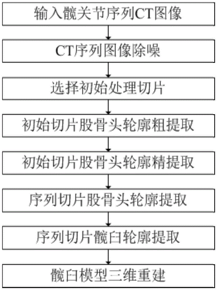
[0063] Example 1, such as figure 1 A method for reconstructing an acetabular tissue model oriented to serialized hip joint CT images as shown, comprising the following steps:

[0064] CT image preprocessing: 3D Gaussian blur method is used to denoise each CT slice in the CT image sequence.

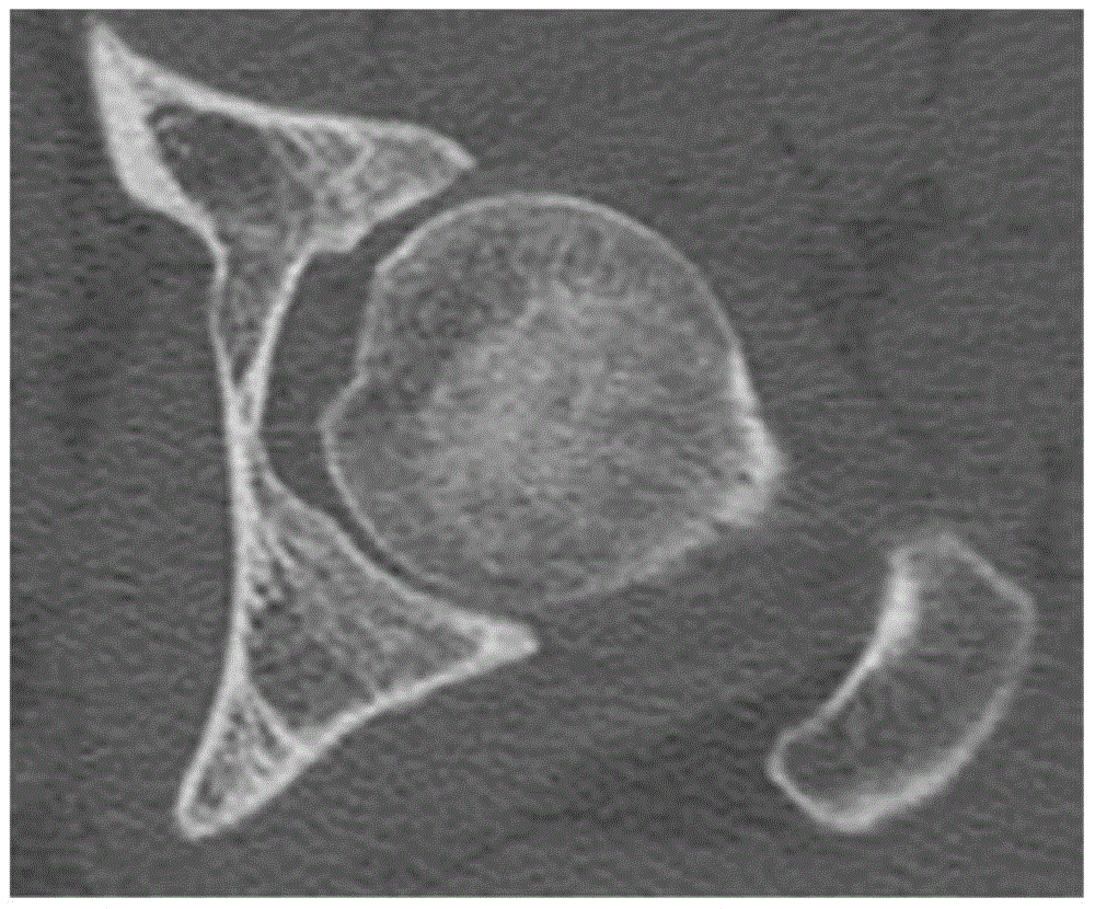
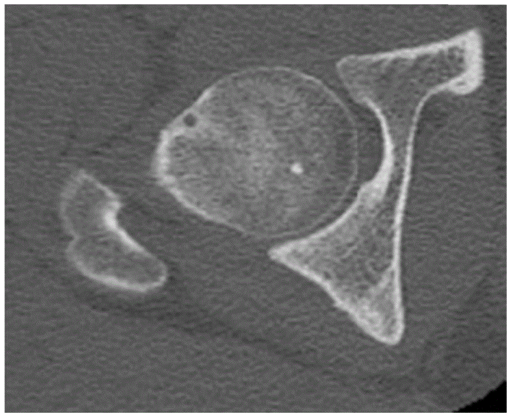
[0065] Select the initial processing slice: the CT slice where the greater trochanter and femoral head were separated for the first time was used as the initial CT slice. Such as figure 2 Shown is an image slice of the left hip joint, image 3 Shown is an image slice of the right hip.

[0066] Create a space Cartesian coordinate system:

[0067] Take the upper left corner of the first slice in the CT image sequence as the coordinate origin, take the rightward direction as the positive direction of the x-axis, take the downward direction as the positive direction of the y-axis, and take the direction in which the number of slice layers increases as the positive direction of the z-axis ...


PUM


Abstract

The invention discloses an acetabulum tissue model reconstruction method for a serialization hip joint CT image. The method includes the following steps of forming a fine outline, wherein the point with the maximum gradient on the vertical line of the tangent line of any point on a circular caput femoris coarse outline on a selected original CT slice is selected; traversing the caput femoris coarse outline to obtain an outline point set of the points, with the maximum gradient, on the vertical lines of all the tangent lines; connecting the points in the outline point set to form the fine outline of the caput femoris tissue of the CT slice; extracting a sequence caput femoris outline; extracting a sequence acetabulum image; conducting three-dimensional reconstruction. According to the method, personalized related position parameters are obtained on the basis of the individual hip joint bone shape of a patient, acetabulum segmentation is conducted in the CT image to obtain accurate acetabulum tissue images and a three-dimensional model, the foundation is laid for conducting personalized reverse determination modeling on an artificial caput femoris prosthesis in the subsequent process, no priori knowledge obtained by conducting training on other data sets is needed, and implementation is easy.

Description

technical field [0001] The invention relates to a method for reconstructing an acetabular tissue model, in particular to a method for reconstructing an acetabular tissue model oriented to serialized CT images of the hip joint. Involving patent classification A61 Medicine or veterinary medicine; Hygiene A61B Diagnosis; Surgery; Appraisal A61B6 / 00 Apparatus for radiodiagnosis, such as A61B6 / 02 Apparatus for diagnosis in different planes combined with radiotherapy equipment in sequence; Stereoradiography Diagnostic A61B6 / 03 computerized tomography camera. Background technique [0002] In the hemihip replacement clinical operation for the treatment of femoral head necrosis, the shape of the acetabular bone varies greatly in different patients. If a standard artificial femoral head prosthesis is used, the postoperative effect is not ideal. Personalized design is very necessary. The following technical solutions are now adopted to solve this problem; [0003] Technology 1: Segm...

Claims


Application Information

Patent Timeline
Patent Type & Authority Applications(China)
IPC IPC(8): G06T17/00
Inventor 刘斌张辉
Owner DALIAN UNIV OF TECH
Learn More