
Method for rapidly selecting and displaying interested blood vessel in magnetic resonance blood vessel image

A technology for rapid selection and blood vessel images, applied in image analysis, image data processing, instruments, etc., can solve the problems of complex calculation of blood vessel models, time-consuming, inaccurate detection of small blood vessels in branches, etc., to improve the efficiency of clinical diagnosis , reduce the time required, solve the effect of manual segmentation problems

Inactive Publication Date: 2014-06-25
EAST CHINA NORMAL UNIVERSITY
View PDF3 Cites 29 Cited by
  • Summary
  • Abstract
  • Description
  • Claims
  • Application Information

AI Technical Summary

Problems solved by technology

[0005] The present invention overcomes the defects of relatively large calculation complexity of the vessel model in the prior art, long time-consuming and inaccurate detection of branched small vessels, and proposes a method for quickly selecting and displaying vessels of interest in magnetic resonance vessel images

Method used


Image

  • Method for rapidly selecting and displaying interested blood vessel in magnetic resonance blood vessel image
  • Method for rapidly selecting and displaying interested blood vessel in magnetic resonance blood vessel image
  • Method for rapidly selecting and displaying interested blood vessel in magnetic resonance blood vessel image

Examples


Embodiment 1

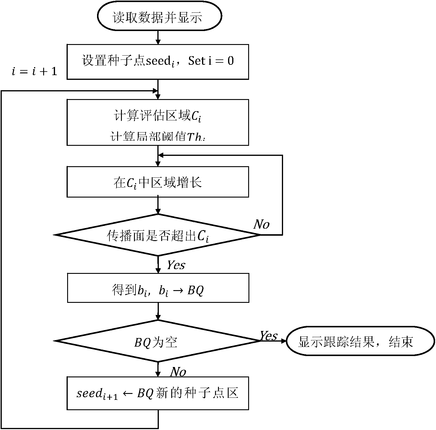
[0061] The downstream vessel selection technology adopts the directional region growing method based on adaptive threshold as the core algorithm, Figure 7 Shown is a schematic diagram of the distribution of the evaluated areas during growth. The specific implementation steps are as follows:

[0062] First determine the starting point and downstream direction. This step is completed in the user interaction step S2, and the user can directly specify the starting point and downstream direction of blood vessel tracking on the three-dimensional image.

[0063] Compute local evaluation regions and local thresholds. After the starting point is determined, the algorithm will start to calculate the size and position of the local evaluation area with the starting point as the center point, and use the iterative optimization threshold detection method to calculate the local threshold in the local evaluation area. The local threshold is used To distinguish background signal from vascu...

Embodiment 2

[0075] The same segmentation algorithm is also used in the selection technology of connected blood vessels between two points. The difference is that in the directional selection of the directional region growth, the local blood vessel segmentation technology uses the minimum path algorithm in the direction selection, and if the growth direction of the directional region is far away from the target point in the Euclidean distance, try other candidate directions for orientation. Region growing, so that it can quickly track from the starting point to the end point, the specific implementation steps are as follows:

[0076] First determine the starting point and downstream direction. This step is completed in the user interaction step S2, and the user can directly designate the starting point and the ending point of blood vessel tracking on the three-dimensional image.

[0077] After the starting point is determined, the algorithm starts to calculate the size and position of the...


PUM


Abstract

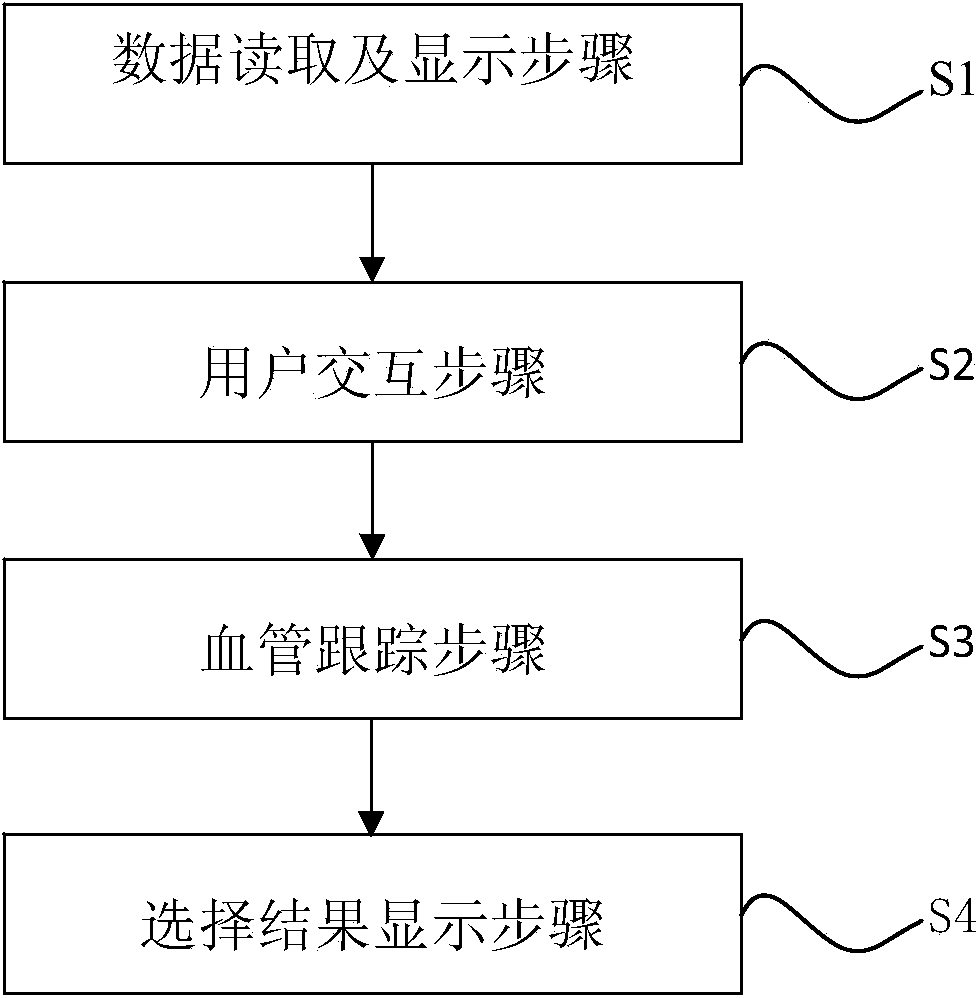
The invention discloses a method for rapidly selecting and displaying an interested blood vessel in a magnetic resonance blood vessel image. The method comprises the following steps that data are read and displayed, and data of an original image are loaded and the original image is displayed on a terminal; users interact with each other, a starting point is selected in interested areas of the blood vessel selected from the original image, and the growth direction of the blood vessel is determined; the blood vessel is tracked, orientation area growth is carried out from the starting point in the growth direction of the blood vessel to obtain part of the intersected blood vessel areas until all the intersected blood vessel areas are obtained; the selection result is displayed, all the areas of the interested blood vessel are displayed in a three-dimensional rebuilding process. Through the method for rapidly selecting and displaying the interested blood vessel in the magnetic resonance blood vessel image, the interested blood vessel in the magnetic resonance blood vessel image can be rapidly, stably and practically selected.

Description

technical field [0001] The invention belongs to the technical field of magnetic resonance imaging, in particular to a method for quickly selecting and displaying interested blood vessels in magnetic resonance blood vessel images. Background technique [0002] Magnetic resonance imaging has the advantages of no damage, high soft tissue contrast, and slices in any direction. It can not only provide tissue morphological information, but also reflect tissue functional characteristics and molecular level information. Therefore, magnetic resonance imaging has been widely used in medical clinical diagnosis. Magnetic resonance angiography also plays an important role in the field of vascular imaging. [0003] Although three-dimensional magnetic resonance angiography has many advantages, it also has its own defects: 1. It can display the whole intracranial blood vessels at the same time, and cannot target the blood vessels of interest, and small lesions are easily obscured by other b...

Claims


Application Information

Patent Timeline
IPC IPC(8): G06K9/32G06T7/00
Inventor 王成龙谢海滨宁瑞鹏杨光
Owner EAST CHINA NORMAL UNIVERSITY
Learn More