Immunohistochemical diagnosis kit for colorectal cancer malignancy and metastasis of colorectal cancer
A diagnostic kit and technology for colorectal cancer, which can be used in disease diagnosis, biological testing, material testing, etc., and can solve the problems of lack of experiments and research
- Summary
- Abstract
- Description
- Claims
- Application Information
AI Technical Summary
Problems solved by technology
Method used
Image
Examples
Embodiment 1
[0023] Example 1 Preparation of Immunohistochemical Diagnostic Kit for Malignancy and Metastasis of Colorectal Cancer
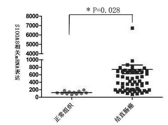
[0024] The inventor used the NCBI high-throughput gene expression database GEO DataSets (gene expression omnibus) to analyze the S100A8 RNA level microarray data of 70 cases of colorectal cancer tissues and 12 cases of normal colorectal tissues in the series GSE9348. The results are shown in the attached figure 1 shown. Through this study, it was found that the expression level of S100A8 in colorectal cancer was significantly higher than that in normal colorectal tissue, p<0.05, and the difference was statistically significant. Further experiments by the inventors by immunohistochemistry showed that the expression level of S100A8 in colorectal cancer was significantly higher than that in the normal colorectal group, and the expression level of S100A8 protein was significantly different in colorectal cancer tissues with different degrees of differentiation, hi...
Embodiment 2
[0028] Example 2 Using the kit prepared in Example 1 to detect colorectal cancer tissue samples
[0029] (1) Dewaxing of baked slices: Take the tissues of colorectal cancer patients, put the tissue slices to be stained in a 60°C oven and bake for 60-90 minutes (depending on the quality of the slices), then quickly put them in xylene (I, II ) for 10 minutes × 2 times for dewaxing;
[0030] (2) Hydration: Place the slices in the following gradient alcohols in order: soak in 100% ethanol for 5 minutes - soak in 90% ethanol for 5 minutes - soak in 75% ethanol for 5 minutes - soak in 50% ethanol for 5 minutes -- soak in distilled water for 3 minutes -- wash with PBS for 3 minutes x 3 times;
[0031] (3) Antigen retrieval: Boil 0.01M citric acid antigen retrieval solution at pH6.0 in a microwave oven, place slices in it, seal the container mouth with latex gloves, continue heating in boiling water for 15-20 minutes, and keep the temperature at 100°C , After the container is natura...
Embodiment 3
[0041] Example 3 Using the kit of the present invention to detect colorectal cancer tissue samples for clinical verification
[0042] The expression of S100A8 protein in 54 cases of normal and 104 cases of colorectal cancer gross specimens was detected by using the kit of Example 1 and according to the method and steps of Example 2. All tissue samples were pathologically confirmed by the Pathology Department of the Second Xiangya Hospital of Central South University after surgery; 58 of the 104 colorectal tumor samples were non-metastatic cancers, and 46 were distant metastatic cancers; 96 of them could be used for malignancy analysis: There were 14 well-differentiated carcinomas, 48 moderately differentiated carcinomas, and 34 poorly differentiated carcinomas.
[0043] According to the localization of the protein in the cell, a comprehensive score was made according to the two criteria of protein expression positive signal intensity and positive expression cell number. Cri...
PUM
 Abstract
Description
Claims
Application Information
 - R&D
- Intellectual Property
- Life Sciences
- Materials
- Tech Scout
- Unparalleled Data Quality
- Higher Quality Content
- 60% Fewer Hallucinations
Browse by: Latest US Patents, China's latest patents, Technical Efficacy Thesaurus, Application Domain, Technology Topic, Popular Technical Reports.
© 2025 PatSnap. All rights reserved.Legal|Privacy policy|Modern Slavery Act Transparency Statement|Sitemap|About US| Contact US: help@patsnap.com


