
Cancer cell or other pathologic cell detection diagnostic device

A technology of pathological cells and diagnostic devices, applied in the field of diagnostic instruments, can solve the problems of limited application, inability to dynamically monitor changes in the treatment process of tumor cells, tumor cell metastasis, etc., and achieve the effect of wide application, light weight and fast speed

Active Publication Date: 2013-10-23
广州睿辰生物科技有限公司
View PDF4 Cites 6 Cited by
  • Summary
  • Abstract
  • Description
  • Claims
  • Application Information

AI Technical Summary

Problems solved by technology

However, most of the current applications are still in the research stage, especially the basis of the diffusion mechanism under living conditions needs to be further studied, and its clinical application value needs to be further explored, and more experience needs to be accumulated before it is widely used in clinical practice.
[0008] Although tumor diagnosis methods such as gene diagnosis, radioimmunoimaging, molecular nuclear medicine technology, and magnetic resonance diffusion weighted imaging have been developed, these methods are mainly used for static qualitative analysis, and cannot dynamically monitor the changes of tumor cells during treatment and Tumor cell metastasis limits its application in basic research and clinical practice, especially in the accurate quantitative analysis of cell numbers required in the diagnosis and treatment of major diseases such as malignant tumors

Method used


Image

  • Cancer cell or other pathologic cell detection diagnostic device
  • Cancer cell or other pathologic cell detection diagnostic device
  • Cancer cell or other pathologic cell detection diagnostic device

Examples


Embodiment Construction

[0049] The specific implementation manner of the present invention will be described in further detail below by describing the best embodiment with reference to the accompanying drawings.

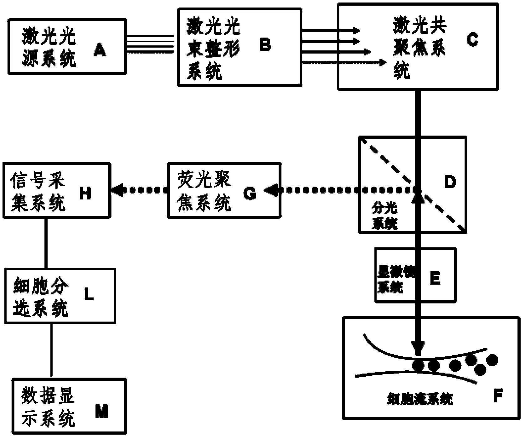
[0050] Such as figure 1 As shown, the tumor cell or other pathological cell detection and diagnosis device consists of laser light source system A (excitation light), laser beam shaping system B, laser confocal system C, spectroscopic system D, microscope system E, cell flow system or cell collection System F, fluorescence focusing system G, signal acquisition system H, cell sorting system L, data display system M and other parts; among them, signal acquisition system H, cell sorting system L and data display system M constitute the cell analysis system; signal acquisition System H is a photoelectric converter that can convert optical signals into electrical signals. Cell sorting system L and data display system M constitute the control system, and signal acquisition system H is connected t...


PUM


Abstract

The invention discloses a cancer cell or other pathologic cell detection diagnostic device which is composed of a laser source system, a beam shaping system, a laser confocal system, a beam splitting system, a microscope system, a cell flow system or a cell acquisition system, a fluorescence focusing system and a cell analysis system. The laser source system emits laser with different wavelengths and the laser is shaped by the beam shaping system; the focuses are gathered on the same plane by the laser confocal system after the laser is shaped; then the laser enters the microscope system through the beam splitting system and is irradiated on stained cells in the cell flow system or the cell acquisition system; the fluorescence generated by the stained cells after irradiation of the laser is amplified by the microscope system and then enters the beam splitting system; and the amplified fluorescence is reflected to the fluorescence focusing system by the beam splitting system and thereafter enters the cell analysis system for cell separation to obtain separation results. The cancer cell or other pathologic cell detection diagnostic device disclosed by the invention is high in detection speed and high in precision, and can be applied to early diagnosis, early treatment and prognosis of cancer cells and other pathologic cells.

Description

technical field [0001] The invention relates to a diagnostic instrument, in particular to a tumor cell or other pathological cell detection and diagnosis device. Background technique [0002] Conquering cancer has always been a major issue that needs to be solved urgently in the medical field. According to a recent report by the United Nations World Health Organization (WHO), the number of people who die from cancer in the world every year has exceeded "10 million people". Although people pay more and more attention to the early diagnosis of tumors, the 5-year survival rate of cancer patients is still not ideal. According to the current medical level, about 80%-90% of early cancer patients without metastasis can be cured; but for advanced cancer patients, there are fewer people who can survive for more than 5 years after treatment. The fundamental reason is the lack of means to detect and treat tumor tissue at an early or even super early stage. If cancer patients can be d...

Claims


Application Information

Patent Timeline
IPC IPC(8): C12M1/36C12M1/34
Inventor 周辉
Owner 广州睿辰生物科技有限公司
Learn More