Western blot analytical technique
A technique for blotting and analyzing results, applied in the field of western blotting analysis technology, which can solve problems such as errors, expensive photographic film processing components, and difficult maintenance
- Summary
- Abstract
- Description
- Claims
- Application Information
AI Technical Summary
Problems solved by technology
Method used
Image
Examples
example 1
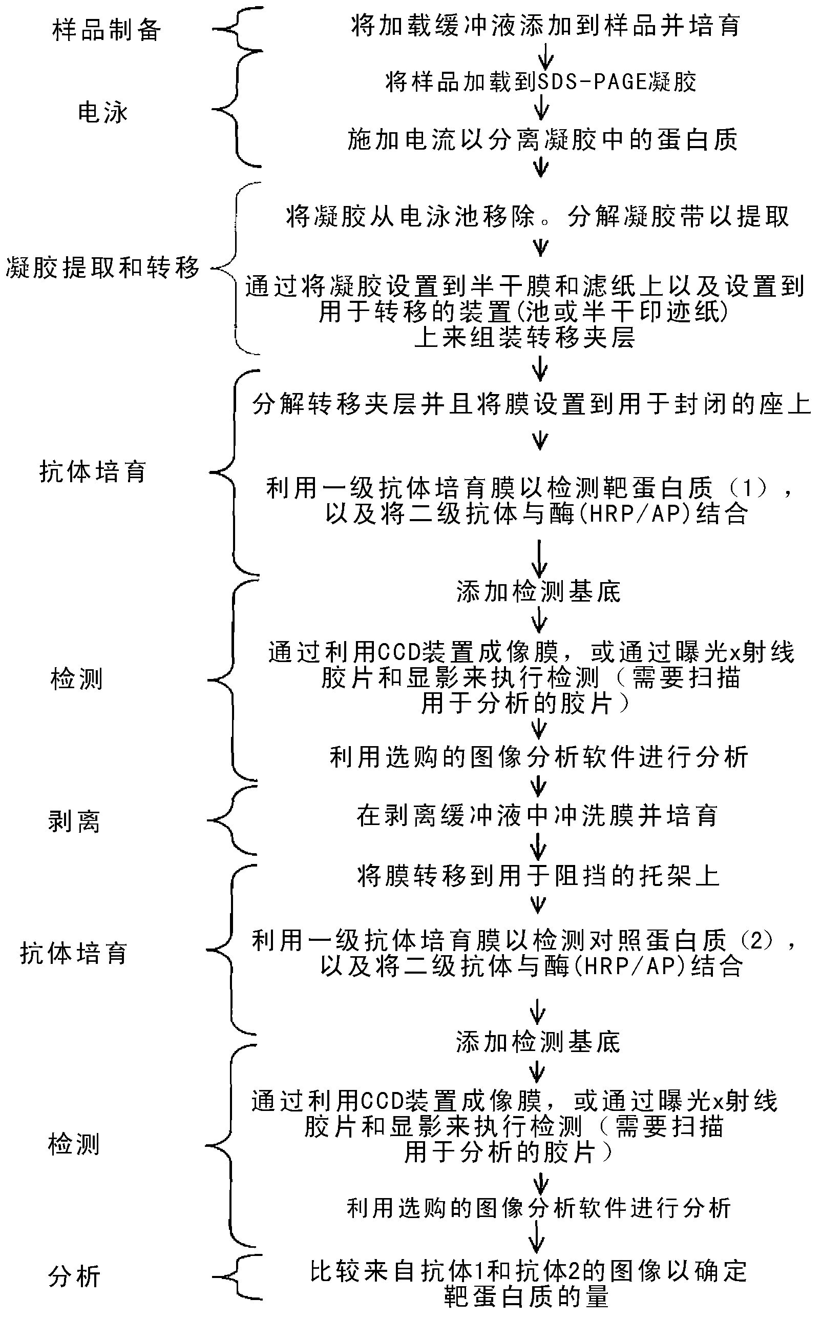
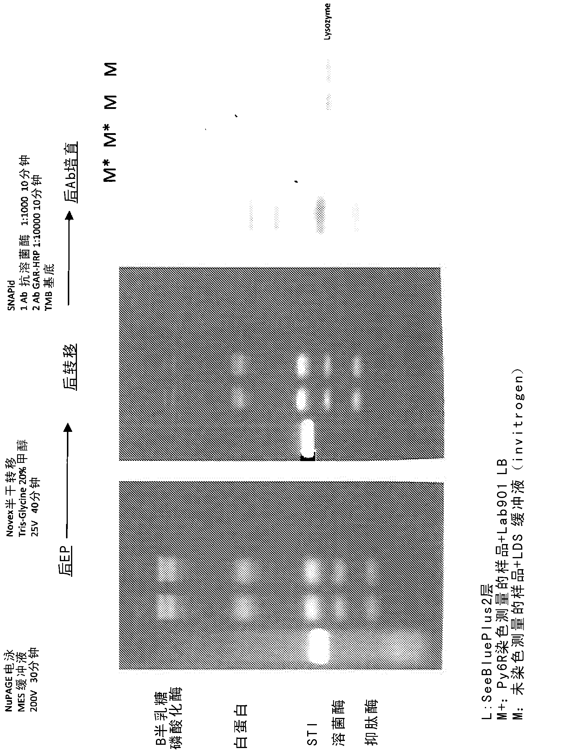
[0085] gel electrophoresis
[0086] Prepare protein-containing samples as follows:
[0087] a. Incubate 20 μl protein sample with 20 μl fluorescent stain for 7 minutes at 75°C; and
[0088] b. Add 40 [mu]l loading buffer, mix and incubate at 75[deg.]C for an additional 5 minutes.
[0089] Additionally, control samples were prepared as follows:
[0090] a. Mix 20 μl protein sample with 10 μl reducing agent and 4x25 μl Invitrogen's LDS buffer; and
[0091] b. Incubate at 75°C for 5 minutes.
[0092] Both samples were loaded onto a NuPAGE electrophoresis gel and run according to the manufacturer's standard protocol to separate the proteins, and an image of the gel was then captured using a UV-transmitter and digital camera.
[0093] Transfer the separated sample to the membrane
[0094] The gel consisted of separated proteins divided into two equal parts and then one half was transferred to a PVDF membrane and the other half to a nitrocellulose membrane. Separated prote...
example 2
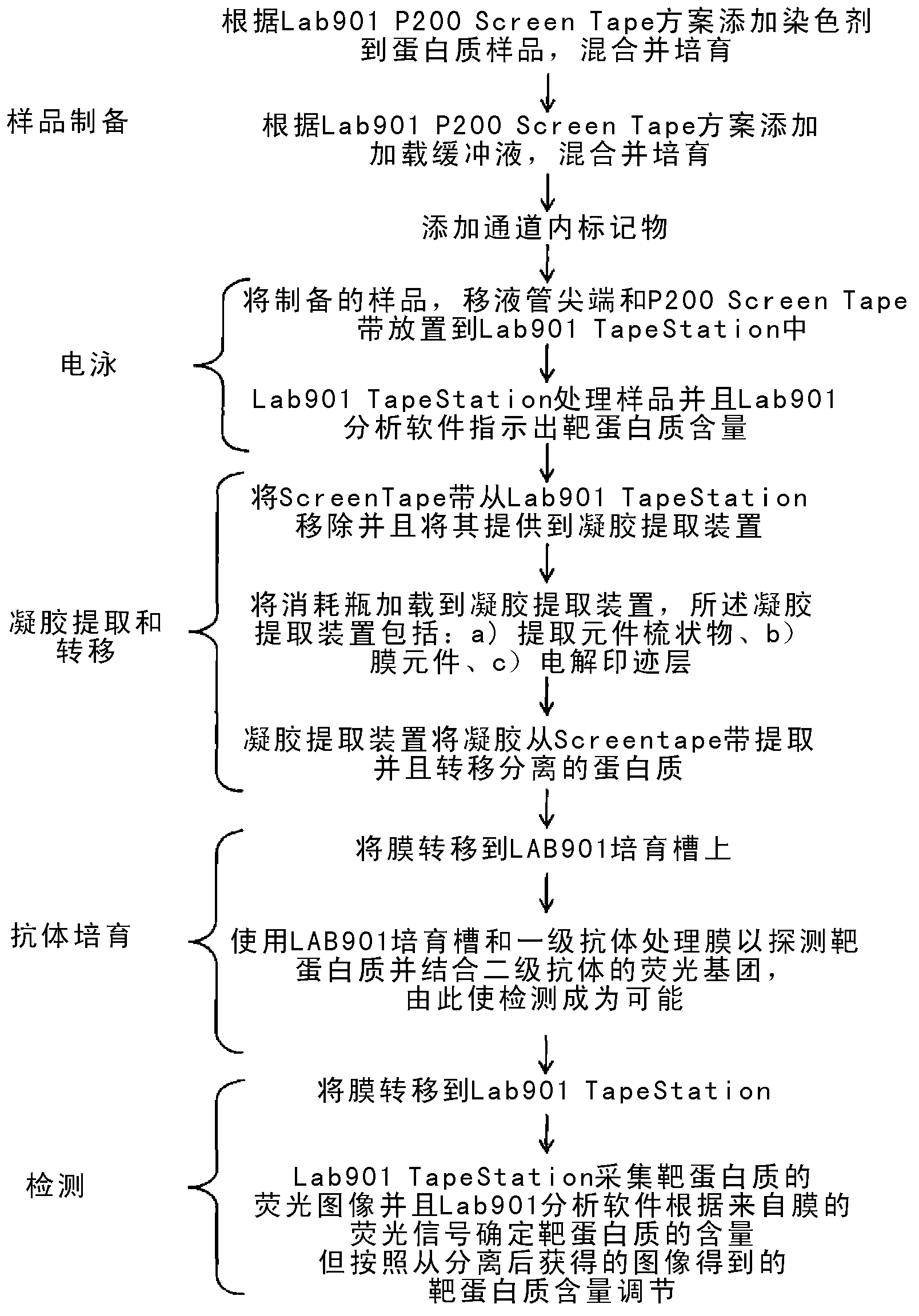
[0108] gel electrophoresis
[0109] Prepare protein-containing samples as follows:
[0110] a. Incubate 2 μl protein sample with 2 μl fluorescent stain for 7 minutes at 75°C;
[0111] b. Add 4 μl of loading buffer, mix and incubate at 75°C for 5 minutes; and
[0112] c. Add 2 μl of in-channel marker.
[0113] Load samples into Lab901P200 Electrophoresis gels were run according to the manufacturer's standard protocol to separate proteins. Use Lab901 for the used imaging (see Figure 5 "P200" in a).
[0114] Transfer the separated sample to the membrane
[0115] including isolated proteins From To recover, remove its carrier layer and use two blades to cut , exposing the top and bottom of the gel column contained within the 16 subcontainers. A comb comprising 16 gel pushing elements was used to push the gel within each sub-container so that the gel was drawn onto a PVDF membrane wetted in 20% methanolic Tris-Glycine transfer buffer. The membrane was placed ...
example 3
[0128] gel electrophoresis
[0129] Prepare protein-containing samples as follows:
[0130] a. Incubate 2 μl protein sample with 2 μl fluorescent stain for 7 minutes at 75°C;
[0131] b. Add 4 μl of loading buffer, mix and incubate at 75°C for 5 minutes; and
[0132] c. Add 2 μl of in-channel marker. Load samples into Lab901P200 Electrophoresis gels were run according to the manufacturer's standard protocol to separate proteins. Use Lab901 (see Figure 6 "P200" in a) used for imaging
[0133] Transfer the separated sample to the membrane
[0134] including isolated proteins From To recover, remove its carrier layer and use two blades to cut , exposing the top and bottom of the gel column contained within the 16 subcontainers. A comb comprising 16 gel pushing elements was used to push the gel within each sub-container so that the gel was drawn onto a PVDF membrane wetted in 20% methanolic Tris-Glycine transfer buffer. Membranes with a single protein transf...
PUM
 Abstract
Description
Claims
Application Information
 - R&D
- Intellectual Property
- Life Sciences
- Materials
- Tech Scout
- Unparalleled Data Quality
- Higher Quality Content
- 60% Fewer Hallucinations
Browse by: Latest US Patents, China's latest patents, Technical Efficacy Thesaurus, Application Domain, Technology Topic, Popular Technical Reports.
© 2025 PatSnap. All rights reserved.Legal|Privacy policy|Modern Slavery Act Transparency Statement|Sitemap|About US| Contact US: help@patsnap.com


