
Positron emission computer tomography system

A positron emission and tomography technology, applied in the field of medical devices, can solve problems such as limiting the application scope of PET, and achieve the effect of reducing scanning cost, fast scanning speed, and broadening application scope

Inactive Publication Date: 2012-12-12
BEIJING TOP GRADE HEALTHCARE MEDICAL EQUIP CO LTD
View PDF3 Cites 16 Cited by
  • Summary
  • Abstract
  • Description
  • Claims
  • Application Information

AI Technical Summary

Problems solved by technology

However, it takes more than 9 minutes to scan a body position with 32-ring PET, and it takes about an hour to scan a whole body with six body positions. In this way, some elements with short half-lives cannot be used as markers, which limits the application range of PET.

Method used


Image

  • Positron emission computer tomography system
  • Positron emission computer tomography system
  • Positron emission computer tomography system

Examples


Embodiment Construction

[0039] In order to make the technical problems, technical solutions and advantages to be solved by the embodiments of the present invention clearer, the following will describe in detail with reference to the drawings and specific embodiments.

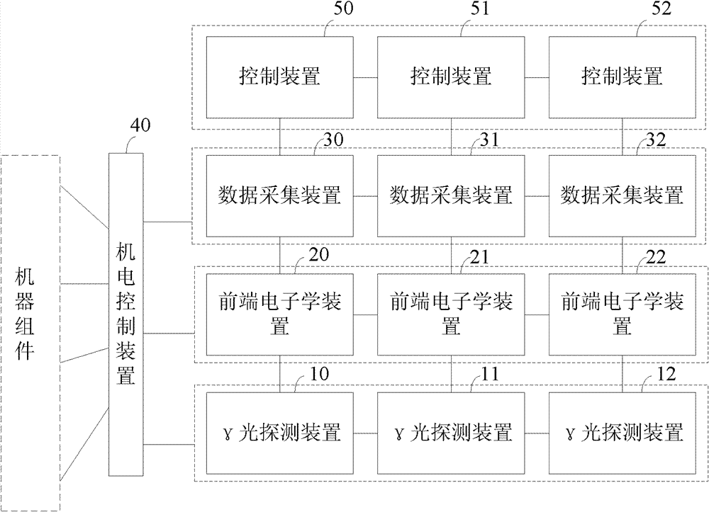
[0040] Embodiments of the present invention aim at the time-consuming problem of using a 32-ring positron emission computed tomography (PET) for scanning in the prior art, and provide a positron emission computed tomography system that can Increase the scanning speed, thereby broadening the application range of PET.

[0041] figure 1 It is a structural schematic diagram of a positron emission computed tomography system according to an embodiment of the present invention, as figure 1 As shown, this embodiment includes:

[0042] Three gamma light detection devices 10, 11 and 12 are respectively used to generate gamma photons that hit the crystal to generate visible light, and convert the visible light into electrical signals;

[0043]...


PUM


Abstract

The invention provides a positron emission computer tomography system and belongs to the field of medical devices. The system comprises three gamma-ray detection devices, three front-end electronics devices, three data acquisition devices, an electromechanical control device and three control devices, wherein three gamma-ray detection devices are used for generating gamma photons to hit crystals to produce visible light and converting the visible light into electric signals; three front-end electronics devices are used for determining single case information according to electric signals of corresponding gamma-ray detection devices; three data acquisition devices are used for acquiring the single case information obtained by corresponding front-end electronics devices and processing the single case information to obtain coincidence case information; the electromechanical control device is connected with data acquisition devices and is used for controlling movement of machine assemblies under the control of data acquisition devices; and three control devices which are correspondingly connected with data acquisition devices one by one are used for receiving the coincidence case information transmitted by corresponding data acquisition devices, and positron emission tomography (PET) images are obtained through processing. By the aid of the positron emission computer tomography system, the scanning speed can be accelerated, and the application range of PET can be broadened.

Description

technical field [0001] The invention relates to the field of medical equipment, in particular to a positron emission computed tomography system. Background technique [0002] Positron emission computed tomography (PET) is an instrument specially designed to detect the radiation of positron-emitting radionuclides in the body and perform tomographic imaging. Imaging of organs and lesions based on differences in radioactive concentrations of imaging agents between lesions. [0003] During the diagnosis, the tracer drug labeled with nuclide that emits positrons is injected into the human body, the positrons emitted by the nuclide combine with the electrons in the human body, and the γ-rays produced by annihilation pass through the human body, measured by PET in vitro, and recorded by the instrument The intensity of consistent rays passing through the human body, through which the distribution of positrons in the human body can be known and scanned for imaging. [0004] PET con...

Claims


Application Information

Patent Timeline
Patent Type & Authority Applications(China)
IPC IPC(8): A61B6/02
Inventor 孙启银刘小平翟巧波
Owner BEIJING TOP GRADE HEALTHCARE MEDICAL EQUIP CO LTD
Learn More