Method for fully-automatically segmenting and quantifying left ventricle of cardiac magnetic resonance image

A magnetic resonance image, automatic segmentation technology, applied in diagnostic recording/measurement, medical science, diagnosis, etc., can solve the problem of not being able to automatically identify the bottom and top of the left ventricle, not being able to automatically extract the blood volume at the bottom of the left ventricle, and not being able to automatically accurately and effectively locate problems with left ventricle

Inactive Publication Date: 2012-04-04
平安颖像(嘉兴)软件有限公司 +1
View PDF4 Cites 29 Cited by
  • Summary
  • Abstract
  • Description
  • Claims
  • Application Information

AI Technical Summary

Problems solved by technology

However, these algorithms are almost only effective when segmenting the left ventricle completely surrounded by the myocardium, and there are generally some problems as follows: the left ventricle cannot be accurately and effectively automatically located, the bottom and top of the left ventricle cannot be automatically identified, and the bottom of the left v

Method used


Image

  • Method for fully-automatically segmenting and quantifying left ventricle of cardiac magnetic resonance image
  • Method for fully-automatically segmenting and quantifying left ventricle of cardiac magnetic resonance image
  • Method for fully-automatically segmenting and quantifying left ventricle of cardiac magnetic resonance image

Examples


Embodiment Construction

[0036] The features of the present invention and other related features will be further described in detail below in conjunction with the accompanying drawings through the embodiments:

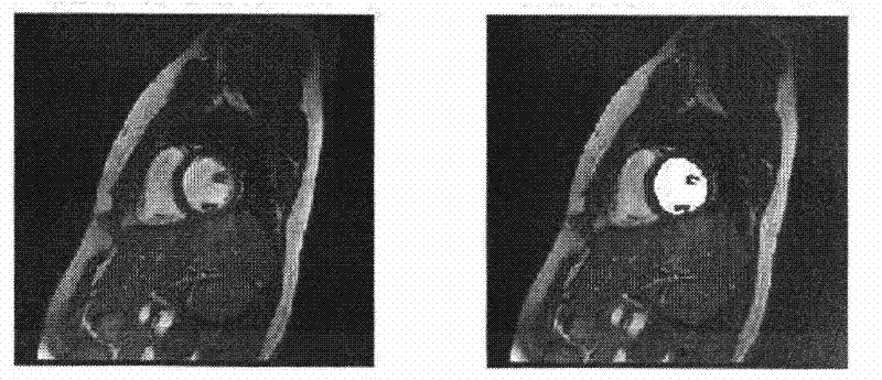
[0037] The method of the present invention accurately and efficiently quantifies the left ventricle function index of the 4D cardiac magnetic resonance image (CMRI) automatically without any manual intervention. The following example introduces step by step the specific operation process of the method of the present invention to automatically locate the left ventricle, automatically determine the top and bottom positions of the left ventricle, and automatically segment and quantify the left ventricle.

[0038] The magnetic resonance imaging data collected in this embodiment is cardiac magnetic resonance imaging data. The data come from GE Signa 1.5T magnetic resonance imaging system, and the imaging sequence selected is SSFP sequence. Specific imaging parameters: TR 3.3-4.5ms, TE 1.1-2.0ms, f...


PUM


Abstract

The invention discloses a method for fully-automatically segmenting and quantifying the left ventricle of a cardiac magnetic resonance image, comprising the following specific steps of: carrying out automatic denoising and edge enhancement processing on a 4D (four-dimensional) cardiac magnetic resonance image first; automatically and preprimarily determining the center of the left ventricle by utilizing Hough transform, and implementing a region growing technology by taking the center of the left ventricle as a starting seed point to provide a left ventricle full-voxel blood region, and taking the mass center of the left ventricle full-voxel blood region as a center of the left ventricle in the current layer; finding out the center of each layer by using a seed propagation technology; implementing a region growing technology based on an iterative falling threshold by taking the center of the left ventricle of each layer as a starting seed point to automatically provide a left ventricle blood region of each layer, and calculating the area of the left ventricle blood region of each layer; automatically segmenting the top of the left ventricle according to the time-space continuity of the area of the left ventricle and calculating the area of the top of the left ventricle; positioning the bottom of the left ventricle according to the area and shape time-space continuity of the left ventricle, and automatically segmenting the bottom of the left ventricle of the heart by adopting a region growing technology which is constricted by the left ventricle shape with time-space continuity, and calculating the area of the bottom of the left ventricle; and finally, realizing whole segmentation of the left ventricle image. The method disclosed by the invention is of a fully-automaticprocess without any manual intervene.

Description

technical field [0001] The present invention belongs to the technical field of magnetic resonance imaging, and specifically refers to a method for automatically and accurately positioning the left ventricle in a 4D cardiac magnetic resonance image, and automatically identifying the bottom and top positions of the left ventricle, thereby realizing fully automatic segmentation and quantification of the left ventricle in the entire cardiac magnetic resonance image method. Background technique [0002] In recent years, heart disease has become the number one killer that endangers human health. In order to improve the quality of life and reduce the mortality rate of heart disease, a large number of technologies have been developed for clinical prospective diagnosis and treatment of heart disease. Accurate evaluation of heart disease with the help of medical imaging technology has become a routine and effective method for clinical diagnosis of heart disease and formulation of tre...

Claims


Application Information

Patent Timeline
IPC IPC(8): A61B5/055
Inventor 王丽嘉裴孟超李建奇
Owner 平安颖像(嘉兴)软件有限公司
Why Eureka
Try Eureka