


Quick Research
Generate reliable direction feasibility study reports for your R&D in just a few steps.

Technical Q&A
Discover and master advanced knowledge NOW. Basics, ideas, possibilities, all at once.

Find Solutions
As an expert in R&D theories, this can generate solutions to your technical problems instantly.

Evaluate Feasibility
Analyze your overall solution with one click, know your potential R&D risks in advance.

Monitor Landscape
Get weekly tech updates, stay abreast of the latest tech innovations and key insights.
Method for manufacturing anthropotomy cast specimen model
A production method and anatomical technology, applied in teaching models, special data processing applications, instruments, etc., can solve the problems of large demand for specimens, high manufacturing costs, and inability to use mold-turning technology
- Summary
- Abstract
- Description
- Claims
- Application Information
AI Technical Summary
Problems solved by technology
Method used
Image


Examples
Embodiment 1
[0028] Embodiment 1: Introduce the process of making a lung cast specimen model based on the lung cast specimen.
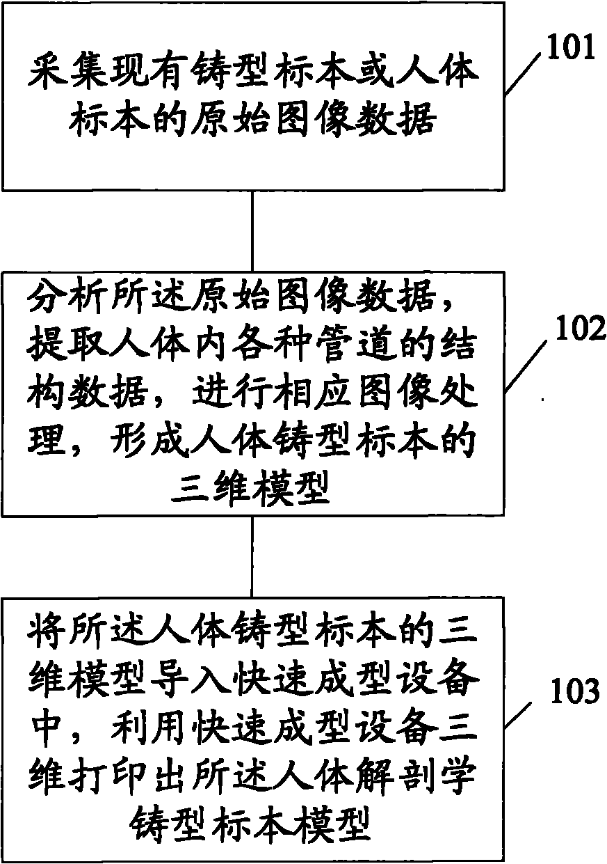
[0029] 1. Perform CT scanning or three-dimensional laser scanning on the lung cast specimen to obtain the tomographic image data of the lung cast specimen.
[0030] 2. Import the continuous tomographic data of the lung cast specimens into the 3D reconstruction software Mimics, integrate the "threshold segmentation", "region growth" and other functional items that come with the 3D reconstruction software, perform image segmentation on the tomographic image data, and manually judge and adjust Two-dimensional images of structures such as pulmonary artery, pulmonary vein, trachea, and bronchi are extracted, and then three-dimensionally reconstructed to obtain three-dimensional computer models of various pipeline structures of lung cast specimens.
[0031] 3. Import the obtained computer 3D model of the lung cast specimen into the image processing software 3-Matic for ...
Embodiment 2
[0033] Embodiment 2: Introduce the process of making a cast specimen model of the calf with bone and artery structure based on the scanning data of the calf with bone and artery structure of the human specimen.
[0034] 1. Carry out CT scanning or three-dimensional laser scanning on the calf of the human specimen to obtain tomographic image data of the calf structure.
[0035] 2. Import the continuous tomographic image data of the calf structure into the 3D reconstruction software Mimics, use different thresholds to perform "threshold segmentation" on the bone and artery structure, and use functions such as two-dimensional image modification and region growth to realize the image of the tomographic image Segment and delete the skin, muscle, nerve tissue, etc. in the image, extract the two-dimensional image of the calf bone and arterial structure, and perform three-dimensional reconstruction on this to obtain the three-dimensional computer model of the calf cast specimen.
[00...
PUM
 Abstract
Description
Claims
Application Information
 - R&D Engineer
- R&D Manager
- IP Professional
- Industry Leading Data Capabilities
- Powerful AI technology
- Patent DNA Extraction
Browse by: Latest US Patents, China's latest patents, Technical Efficacy Thesaurus, Application Domain, Technology Topic, Popular Technical Reports.
© 2024 PatSnap. All rights reserved.Legal|Privacy policy|Modern Slavery Act Transparency Statement|Sitemap|About US| Contact US: help@patsnap.com