Encoding detection method based on polymer microsphere change

A coding detection and detection method technology, applied in the field of biomedical detection, can solve the problems of complex process, prone to leakage, poor stability, etc., and achieve the effects of narrow peak half-peak width, diverse internal structure and small dispersion coefficient of detection spectrum.

Inactive Publication Date: 2010-09-29
TIANJIN UNIV
View PDF6 Cites 7 Cited by
  • Summary
  • Abstract
  • Description
  • Claims
  • Application Information

AI Technical Summary

Problems solved by technology

[0005] At present, the carrier microspheres of many liquid-phase biochips use fluorescent microspheres. Due to the uneven fluorescence loading of the microspheres, poor stability, easy leakage, different fluorescence efficiencies of different fluorescent wavelengths, and the cost of fluorescent dyes is high and the process is complicated. The application in biomedical detection is limited; the requirements for microspheres are high, not only the microspheres are required to be highly uniform, the surface functional groups are enriched, and they can effectively and stably embed small molecules

Method used


Image

  • Encoding detection method based on polymer microsphere change
  • Encoding detection method based on polymer microsphere change
  • Encoding detection method based on polymer microsphere change

Examples


Embodiment 1

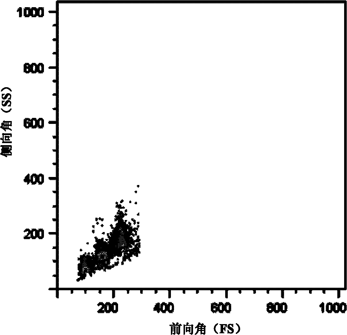
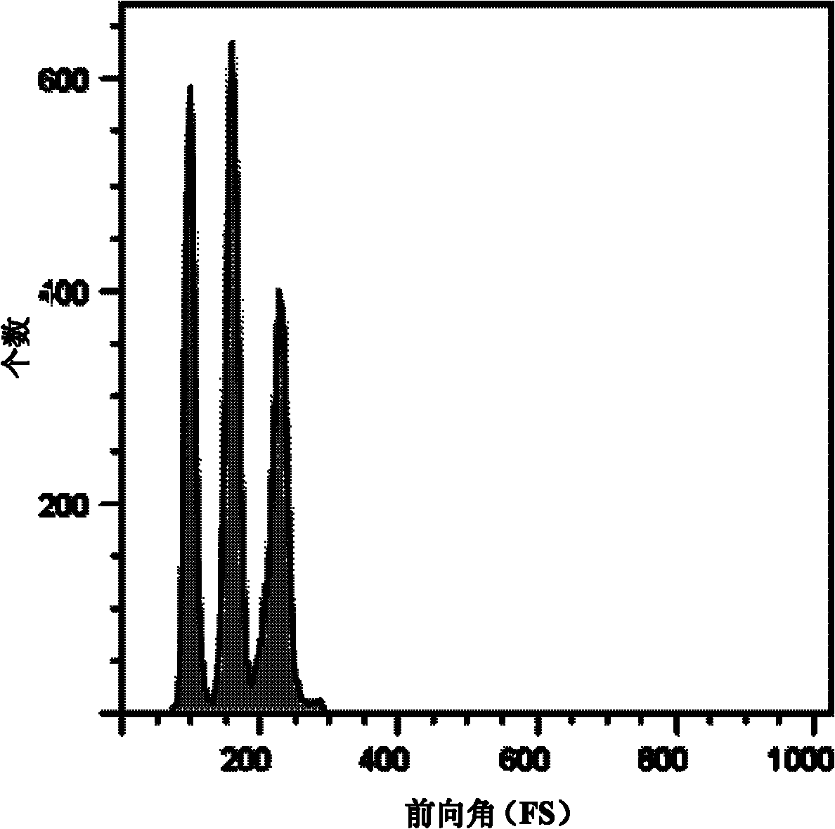
[0035] Select 4μm dense polystyrene-acrylic acid microspheres, the dispersion coefficient is 7.1%, and the surface functional group loading rate is 0.505mmol / g; 5μm dense polystyrene-acrylic microspheres, the dispersion coefficient is 5.0%, and the surface functional group loading The ratio is 0.571mmol / g; 6μm dense polystyrene-acrylic acid microspheres, the dispersion coefficient is 5.5%, and the loading rate of surface functional groups is 0.585mmol / g. Three kinds of microspheres with high cross-linking degree, each 5mg, use 100 μL of PBS buffer was dispersed and then sonicated, labeled as 1, 2, 3. After mixing 1, 2, and 3, ultrasonically disperse and detect with a flow cytometer to obtain a one-dimensional histogram and a two-dimensional scattergram, save and print the results, as shown in Figure 2.

Embodiment 2

[0037] Select 5.7 μm porous polystyrene-acrylic microspheres, the dispersion coefficient is 6.4%, the surface functional group loading rate is 0.597mmol / g, 9.8 μm porous polystyrene-acrylic microspheres, the dispersion coefficient is 5.2% , the surface functional group loading rate is 0.633mmol / g, 12.3μm porous polystyrene-acrylic microspheres, the dispersion coefficient is 7.7%, the surface functional group loading rate is 0.693mmol / g, each 5mg, each with 100μL Disperse in PBS buffer and then sonicate, numbered 1, 2, 3. After mixing 1, 2, and 3, ultrasonically disperse and detect with a flow cytometer to obtain a one-dimensional histogram and a two-dimensional scattergram, save and print the results, as shown in Figure 3.

Embodiment 3

[0039] Select 5.7 μm dense polystyrene-acrylic acid microspheres, the dispersion coefficient is 5.0%, and the surface functional group loading rate is 0, 670 mmol / g; 7.3 μm porous polystyrene-acrylic microspheres, the dispersion coefficient is 5.2%, The loading rate of surface functional groups is 0.633mmol / g; 12.3μm dense polystyrene-acrylic acid microspheres, the dispersion coefficient is 5.5%, and the loading rate of surface functional groups is 0.700mmol / g; 5mg each, buffered with 100μL PBS The liquid is dispersed and then ultrasonicated. The labels are 1, 2, and 3. They are mixed together and dispersed ultrasonically. The flow cytometer is used for detection to obtain a one-dimensional histogram and a two-dimensional scatter diagram. Save and print the results. As shown in Figure 4.

[0040] 2. Detection of active biomolecules


PUM

PropertyMeasurementUnit
particle diameteraaaaaaaaaa
load ratioaaaaaaaaaa

Abstract

The invention relates to an encoding detection method based on polymer microsphere change. The encoding detection method comprises the following steps of: identifying antibody, antigen, DNA or RNA (Ribonucleic Acid) segments in samples to be detected through immunization or gene hybridization specificity by adopting a series of polymer microspheres of the antibody, the antigen, the DNA or the RNA segments with surface coupling specificity, wherein the polymer microspheres have different grain diameters and internal structures; carrying out specific coupling on a molecular probe with the surface carrying fluorescent substances and the antibody, the antigen, the DNA or the RNA segments; detecting fluorescence signals through a detection device, comparing the samples with negative and positive control samples or the samples with known concentration so as to judge whether the samples to be detected are negative or positive; characterizing the grain diameters and the internal structures of the microspheres in the detection device, and accurately judging the substances to be detected according to the corresponding relation between the grain diameters and the internal structures of the microspheres as well as the detection information. The invention is based on immunization and gene hybridization specificity, inaugurates an encoding technology, can realize the detection on a plurality of samples or indexes to be detected simultaneously, and is simple, fast and reliable.

Description

technical field [0001] The invention relates to the technical field of biomedical detection, and mainly relates to a coding detection method based on the change of polymer microspheres. The invention is based on the immune response and gene hybridization specific reaction, innovates the coding technology, and can simultaneously realize the detection of multiple samples or indicators to be tested, which is simple, fast and reliable. Background technique [0002] In the field of biomedicine, people's observation and research on life phenomena have gone deep into the level of single cell and single molecule. Biomedical detection technology is an important means and experimental tool for the development of modern life science and medicine, especially for the clinical diagnosis of infectious diseases, genetic diseases and malignant tumors. [0003] Flow cytometry (Flow Cytometry, FCM) is an analytical method for the quantitative detection and sorting of cells or other biological...

Claims


Application Information

Patent Timeline
Patent Type & Authority Applications(China)
IPC IPC(8): G01N33/53G01N33/533G01N33/576G01N33/569G01N33/571G01N33/68C12Q1/68
Inventor 常津李云红张琦宋涛逯超亮
Owner TIANJIN UNIV
Why Eureka
Try Eureka