Encapsulated endoscope
A capsule-type endoscope and capsule technology, which can be used in endoscopes, radio detectors inside the body, medical science, etc., can solve the problem of not being able to clearly capture large-scale images
- Summary
- Abstract
- Description
- Claims
- Application Information
AI Technical Summary
Problems solved by technology
Method used
Image
Examples
Embodiment approach 1
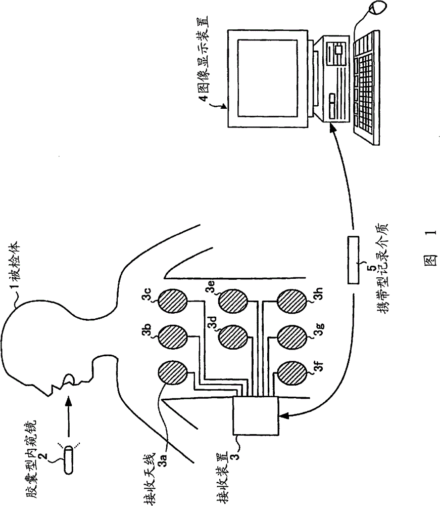
[0044] figure 1 It is a schematic diagram showing a configuration example of an in-vivo information acquisition system including the capsule endoscope according to Embodiment 1 of the present invention. Such as figure 1 As shown, the in-subject information acquisition system includes: a capsule endoscope 2 that captures images inside the subject 1 ; receiving device 3 ; image display device 4 for displaying images of the subject 1 received by receiving device 3 ; and portable recording medium 5 for exchanging data between receiving device 3 and image display device 4 .
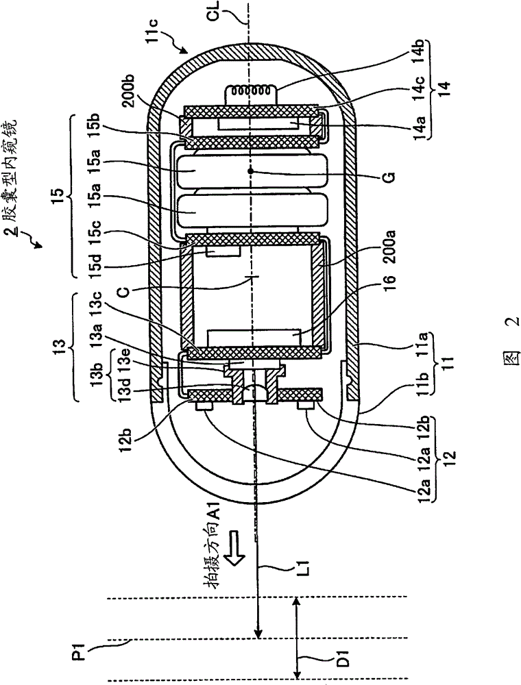
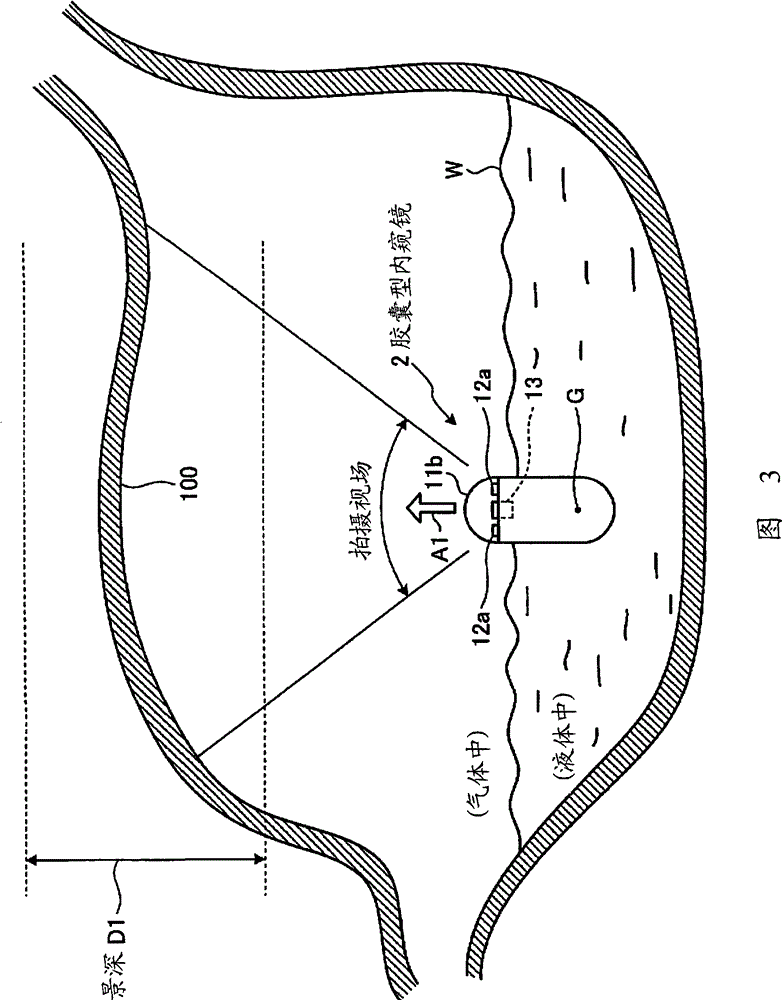
[0045] The capsule endoscope 2 has an imaging function of sequentially capturing images of the inside of the subject 1 in time series and a wireless communication function of sequentially wirelessly transmitting the captured images of the inside of the subject 1 to the outside. Furthermore, the specific gravity of the capsule endoscope 2 is set so that it can float on the surface of a desired liquid such a...
Embodiment approach 2
[0088] Next, Embodiment 2 of the present invention will be described. In the first embodiment described above, one imaging unit 13 is fixedly arranged inside the casing 11 and fixedly arranged on the opposite side of the center of gravity G of the capsule endoscope 2 with the center C of the casing 11 as a boundary. Mode 2 is a multi-eye capsule endoscope in which the imaging unit is fixedly arranged on the opposite side and the same side (centroid side) of the capsule endoscope's center of gravity with the center of the housing as a boundary.
[0089] Figure 4 It is a schematic side sectional view schematically showing a configuration example of the capsule endoscope according to Embodiment 2 of the present invention. Such as Figure 4 As shown, the capsule endoscope 20 of the second embodiment includes a housing 21 instead of the housing 11 of the capsule endoscope 2 of the first embodiment, a control unit 26 instead of the control unit 16, and an illumination unit 22 and...
Embodiment approach 3
[0128] Next, Embodiment 3 of the present invention will be described. In Embodiment 2 above, the center of gravity G of the capsule endoscope 20 is set on the central axis CL of the casing 21, and the imaging direction A1 of the imaging unit 13 is made parallel to the central axis CL of the casing 21. However, in this In Embodiment 3, the center of gravity G of the capsule endoscope is also set at a position separated from the central axis CL, and the direction inclined to the opposite side of the center of gravity G with respect to the central axis CL in the longitudinal direction of the housing is the direction of the imaging unit 13. shooting direction.
[0129] Figure 6 It is a schematic side cross-sectional view schematically showing a configuration example of the capsule endoscope according to Embodiment 3 of the present invention. Such as Figure 6 As shown, the capsule endoscope 30 of the third embodiment has a housing 31 instead of the housing 21 of the capsule en...
PUM
 Abstract
Description
Claims
Application Information
 - R&D
- Intellectual Property
- Life Sciences
- Materials
- Tech Scout
- Unparalleled Data Quality
- Higher Quality Content
- 60% Fewer Hallucinations
Browse by: Latest US Patents, China's latest patents, Technical Efficacy Thesaurus, Application Domain, Technology Topic, Popular Technical Reports.
© 2025 PatSnap. All rights reserved.Legal|Privacy policy|Modern Slavery Act Transparency Statement|Sitemap|About US| Contact US: help@patsnap.com


