
X-ray image apparatus and method of imaging an object under examination

An imaging device, X-ray technology, applied in X-ray equipment, radiation beam guiding devices, instruments for radiological diagnosis, etc. Ensure the effect of meaningful images

Inactive Publication Date: 2009-07-01
KONINKLIJKE PHILIPS ELECTRONICS NV
View PDF2 Cites 11 Cited by
  • Summary
  • Abstract
  • Description
  • Claims
  • Application Information

AI Technical Summary

Problems solved by technology

[0011] However, it remains difficult for a human operator of the system to control to determine whether manually adjusted irradiation parameters are appropriate

Method used


Image

  • X-ray image apparatus and method of imaging an object under examination

Examples


Embodiment Construction

[0058] The figure is shown schematically. In different figures, similar or identical elements are provided with the same reference signs.

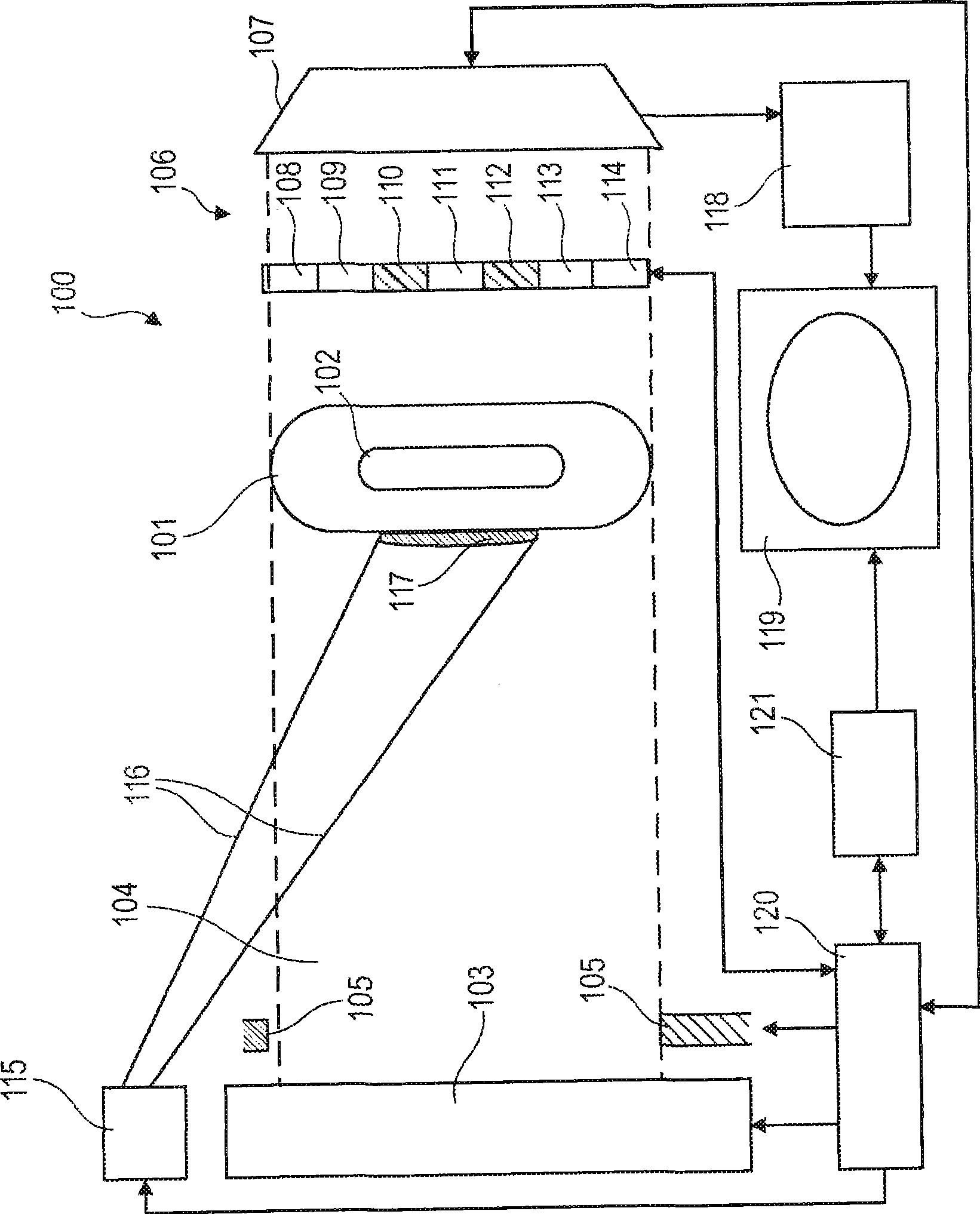
[0059] In the following, refer to figure 1 , an X-ray imaging apparatus 100 according to an exemplary embodiment of the present invention will be described.

[0060] The X-ray imaging apparatus 100 is adapted to image a part of a human body 101 , in the typical case described, a lung 102 .

[0061] The X-ray imaging device 100 comprises an X-ray source 103 (X-ray tube) capable of generating an X-ray beam 104 which is directed towards a human body 101 including in particular a lung portion 102 . The actual width and geometry of beam 104 can be defined using collimator unit 105 .

[0062] Furthermore, a dose measurement device 106 is provided and positioned between the human body 101 and the scintillation detector 107 . The dose measurement device is adapted to spatially correlatively measure the X-ray dose of the X-ray beam 104 in at le...


PUM


Abstract

An X-ray image apparatus (100) for imaging an object under examination (101), the X-ray image apparatus (100) comprising an X-ray source (103) adapted for generating an X-ray beam (104) to be directed to the object under examination (101), a dose measuring device (106) for measuring an X-ray dose of the X-ray beam (104) in at least one selected (110, 112) of a plurality of measurement fields (108 to 114) after transmission of the X-ray beam (104) through the object under examination (102), and an illumination device (115) for illuminating a surface portion (117) of the object under examination (101) which surface portion (117) is indicative of the at least one selected (110, 112) of the plurality of measurement fields (108 to 114).

Description

technical field [0001] The invention relates to an X-ray imaging device. [0002] Furthermore, the invention relates to a method of imaging an examination object. [0003] Among other things, the invention relates to a program element. [0004] Furthermore, the present invention relates to a computer readable medium. Background technique [0005] X-ray imaging is important in many technical fields including medical applications. [0006] Medical imaging is the process by which physicians evaluate areas of a subject's body that are not externally visible. Medical imaging can be clinically motivated to seek diagnosis and examine disease in specific human patients. Alternatively, researchers can use it in order to understand processes going on in living organs. Many techniques developed for medical imaging also have scientific and industrial applications. [0007] In the case of radiography, what is detected is an X-ray beam, which is absorbed at different rates in differ...

Claims


Application Information

Patent Timeline
Patent Type & Authority Applications(China)
IPC IPC(8): A61B6/00G01T1/08H05G1/44
CPCA61B6/542H05G1/26A61B6/508A61B6/08A61B6/00
Inventor L·博格曼J·维歇斯
Owner KONINKLIJKE PHILIPS ELECTRONICS NV
Learn More