Basophilia granulocyte analytical reagent and measuring method
A technology for analyzing basophils and reagents, which is applied in the field of cell analysis reagents, can solve the problems of high price, many measured parameters, complex instrument structure, etc., and achieve the effect of reducing dosage, not easy to separate out, and simple reagent components
- Summary
- Abstract
- Description
- Claims
- Application Information
AI Technical Summary
Problems solved by technology
Method used
Image
Examples
Embodiment 1
[0055] Prepare basophil assay reagents as follows:
[0056] NP40 800mg / L
[0057] Brij 35 2g / L
[0058] Citric acid 10mM
[0059] h 2 O to 1L
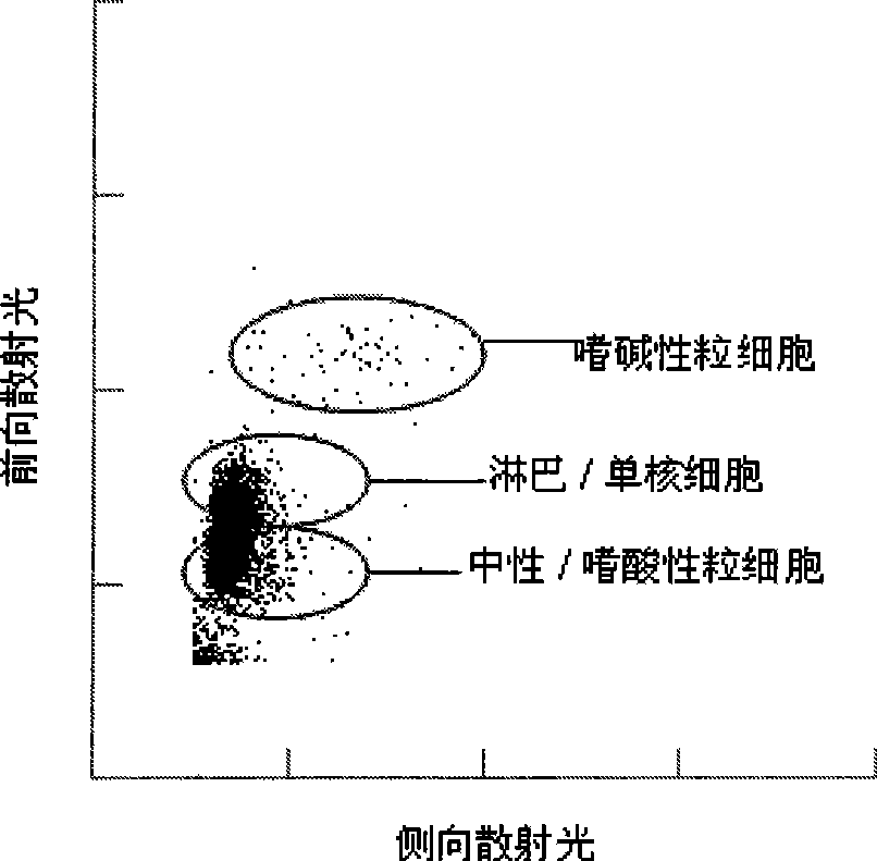
[0060] The pH value of the above reagent is about 2.8. Take 1 mL of the above basophil analysis reagent and mix with 20 μL blood sample, and incubate at 40°C for 10 seconds. Then, the forward and side scattered light signals of the cells were measured on the Mindray BC-5500 hematology analyzer, and the generated scatter diagram was as follows figure 1 . As shown, basophils can be distinguished from other white blood cells using forward and side scatter light signals.
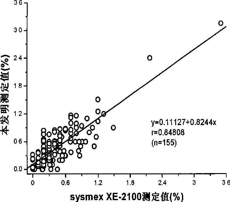
[0061] Another 155 fresh clinical blood samples were taken, and the above-mentioned basophil analysis reagent was used to measure basophils (%) on Mindray BC-5500 blood cell analyzer according to the above method, and the results were compared with the same blood The specimens were analyzed according to the measured values on the sysmex XE-2100 hematology analy...
Embodiment 2
[0070] Prepare basophil assay reagents as follows:
[0071] NP40 800mg / L
[0072] Brij 56 1g / L
[0073] Glycine 20mM
[0074] HCl Amount to adjust pH to 2.5
[0075] h 2 O to 1L
[0076] Using 1 mL of the above-mentioned basophil assay reagent, add 20 μL of blood sample. After incubating at 40°C for 40 seconds, the forward and side scattered light signals of the cells were measured on the Mindray BC-5500 hematology analyzer to generate Figure 4 scatterplot of . As shown, basophils can be classified and counted by forward and side scatter light signals.
Embodiment 3
[0078] Prepare basophil assay reagents as follows:
[0079] Triton X-100 400mg / L
[0080] Brij 35 500mg / L
[0081] Acetic acid 250mM
[0082] h 2 O to 1L
[0083] The pH value of the above reagent is about 2.8. Use 1 mL of the above basophil analysis reagent, mix with 20 μL blood sample, and incubate at 40°C for 10 seconds. Then, the forward and side scattered light signals of the cells were measured on the Mindray BC-5500 hematology analyzer, and the scattergram results were as follows Figure 5 . As shown, basophils can be distinguished from other white blood cells using forward and side scatter light signals.
[0084] Implementation column 4
[0085] Prepare basophil assay reagents as follows:
[0086] Triton X-100 400mg / L
[0087] Brij 56 1g / L
[0088] Potassium hydrogen phthalate 20mM
[0089] HCl Amount to adjust pH to 2.5
[0090] h 2 O to 1L
[0091] Using 1 mL of the above-mentioned basophil assay reagent, add 20 μL of blood sample. After incubating at...
PUM
 Abstract
Description
Claims
Application Information
 - R&D
- Intellectual Property
- Life Sciences
- Materials
- Tech Scout
- Unparalleled Data Quality
- Higher Quality Content
- 60% Fewer Hallucinations
Browse by: Latest US Patents, China's latest patents, Technical Efficacy Thesaurus, Application Domain, Technology Topic, Popular Technical Reports.
© 2025 PatSnap. All rights reserved.Legal|Privacy policy|Modern Slavery Act Transparency Statement|Sitemap|About US| Contact US: help@patsnap.com


