
Cad detection system for multiple organ systems

A technology of organ systems and organs, applied in the field of automated computer inspection of medical imaging, can solve problems such as the ability to reduce interpretation time

Inactive Publication Date: 2009-02-11
CARESTREAM HEALTH INC
View PDF5 Cites 15 Cited by
  • Summary
  • Abstract
  • Description
  • Claims
  • Application Information

AI Technical Summary

Problems solved by technology

thus greatly limiting the ability of such CAD systems to substantially reduce interpretation time

Method used


Image

  • Cad detection system for multiple organ systems
  • Cad detection system for multiple organ systems
  • Cad detection system for multiple organ systems

Examples


Embodiment Construction

[0024] The following is a detailed description of preferred embodiments of the present invention with reference to the accompanying drawings, in each of the several drawings, like reference numerals designate like structural elements.

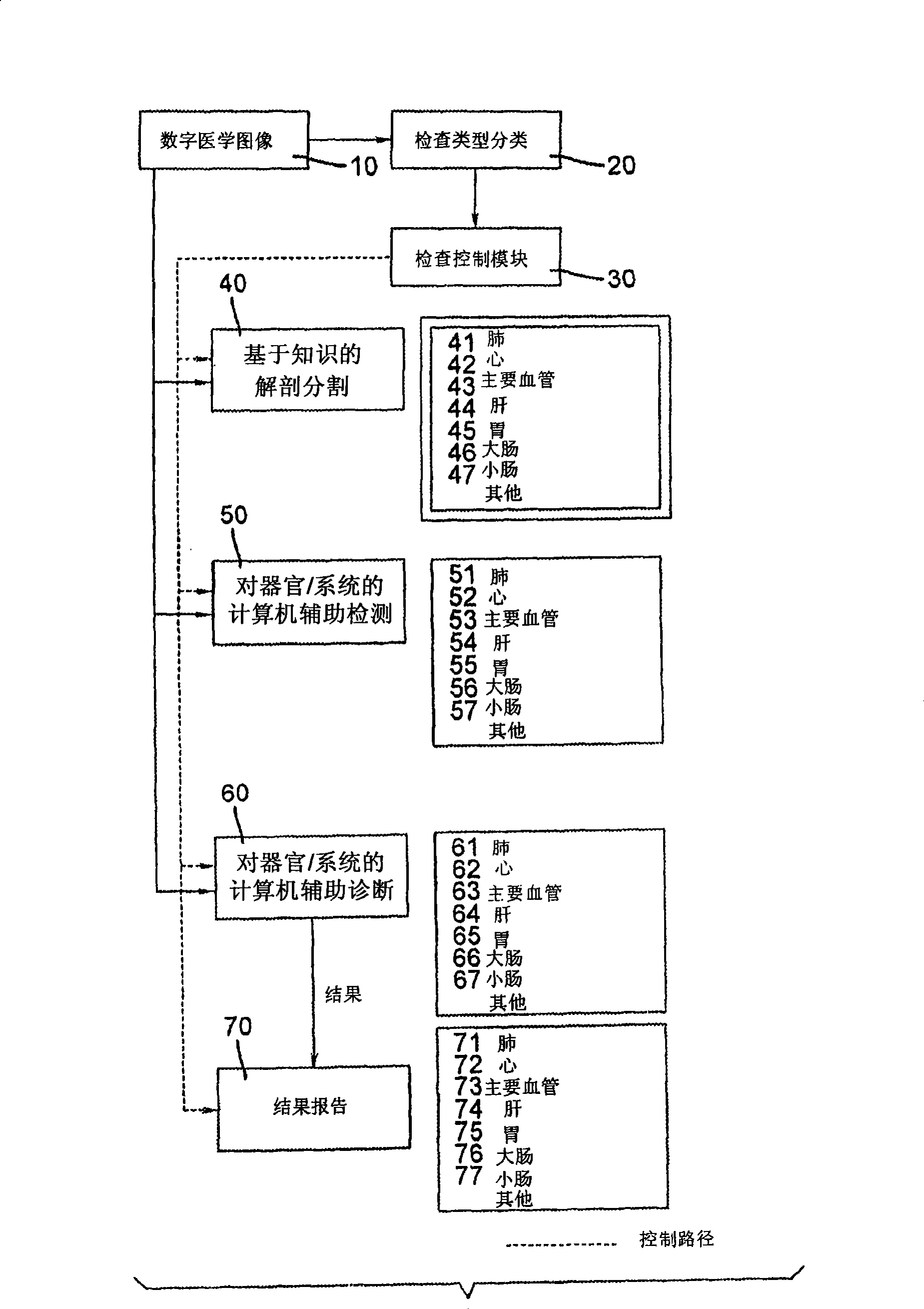
[0025] figure 1 A block diagram of the present invention is shown. Referring to the figure, a digital medical image 10 occurs in the method. The image may be created by any of the known methods currently practiced in the medical field, such as computed tomography. The images are preferably represented in the industry standard DICOM format.

[0026] The first stage of processing includes an optional examination type classification step 20 . The purpose of this step is to determine the specific patient anatomy present in the digital image in order to direct the rest of the processing to the specific organ or organ system present in the image. For example, a CT image of the chest contains information about the patient's lungs, heart, and major...


PUM


Abstract

A computer aided disease detection system and method for multiple organ systems. The method performs computer aided examination of digital medical images. A patient exam type of a digital medical image is determined. Based on the patient exam type, one or more of a plurality of knowledge based anatomical segmentation blocks are invoked, each block performing image segmentation for a single organ or organ system present in the image. Based on the patient exam type, for each successfully segmented organ or organ system, one or more of a plurality of knowledge based computer aided detection blocks are invoked, each block of which is designed to search for and locate potential disease foci in a particular organ or organ system.

Description

technical field [0001] The present invention generally relates to the field of digital medical image processing, and in particular to a method and system for automatic computer inspection of medical images, the method and system for detecting possible disease sites in multiple human organ systems. Background technique [0002] Technological advances in the field of non-invasive imaging of the human body have benefited medical professionals in their ability to detect, diagnose and treat disease, resulting in improved outcomes and reduced morbidity and mortality. Advances in imaging resolution, functionality, and quality often lead to new applications of various imaging modalities that benefit patients and society. However, such advancements incur human and financial costs, particularly in the costs of imaging studies and the necessary specialized medical expertise required to utilize and interpret the resulting images. Over the past decade, three- and four-dimensional modali...

Claims


Application Information

Patent Timeline
Patent Type & Authority Applications(China)
IPC IPC(8): G06T7/00
Inventor H·尼克蓬斯基
Owner CARESTREAM HEALTH INC
Learn More