Methods and apparatus for performing enhanced ultrasound diagnostic breast imaging

A technology of breast imaging and ultrasonic diagnosis, which is applied in the field of medical diagnostic imaging systems, can solve the problem of acoustic contact at the curved edge of the breast, etc.

Inactive Publication Date: 2007-09-05
KONINKLIJKE PHILIPS ELECTRONICS NV
View PDF3 Cites 13 Cited by
  • Summary
  • Abstract
  • Description
  • Claims
  • Application Information

AI Technical Summary

Problems solved by technology

However, this approach does not solve the problem of acoustic contact at the curved edge of the breast

Method used


Image

  • Methods and apparatus for performing enhanced ultrasound diagnostic breast imaging
  • Methods and apparatus for performing enhanced ultrasound diagnostic breast imaging
  • Methods and apparatus for performing enhanced ultrasound diagnostic breast imaging

Examples


Embodiment Construction

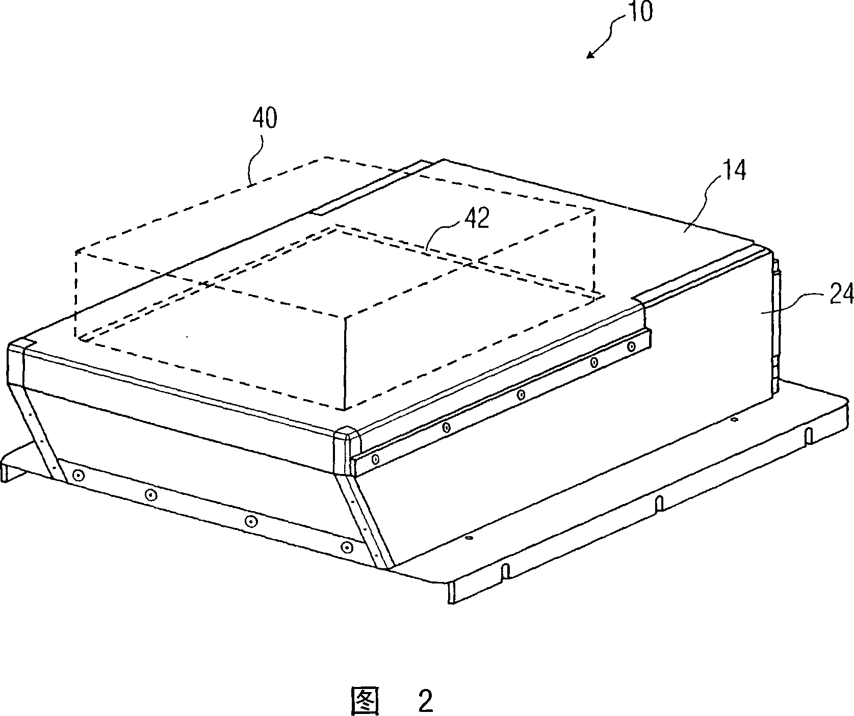
[0016] In the drawings, the same reference numerals denote the same elements. Furthermore, it should be noted that the figures are not drawn to scale.

[0017] Figure 1 is a cross-sectional view of a portion of an ultrasonic diagnostic breast imaging system using a two-pass linear scan to obtain a conventional, unsteered rectangular volume image. That is, the embodiment of FIG. 1 uses unsteered rectangular images to obtain 3D volumes. As a result, a two-pass (A and B) 3D scan with a conventional, non-steering rectangular image frame was unable to image the curved region near the nipple and the tissue adjacent to the chest wall, as described further below.

[0018] As shown in FIG. 1 , an ultrasonic diagnostic breast imaging system 10 includes a first compression plate 12 and a second compression plate 14 . The first and second plates are configured to receive the breast 16 and are further adapted to compress the breast between the first and second plates. Breast 16 extends ...


PUM


Abstract

A method for performing enhanced ultrasound diagnostic breast imaging includes using first and second compression plates (62,64) configured for receiving and compressing a breast between the same. The breast extends from a chest wall (118) of a patient at a proximate end to a nipple at a distal end. Portions of the breast proximate the nipple and proximate lateral edges of the breast are in non-contact with the second compression plate during breast compression. An ultrasound transducer array (68) moves along a path to scan the breast, the ultrasound transducer array being disposed adjacent a side of the second plate (64) opposite the breast. Image data representative of the breast is acquired as the ultrasound transducer array (68) traverses the path. Acquiring image data includes using electronic beam steering with the ultrasound transducer array to acquire image data in either or both (i) a portion (116) of the breast proximate the chest wall and (ii) a portion of the breast in non-contact with the second plate (122).

Description

[0001] Cross-references to related applications [0002] This invention is related to U.S. Patent No. 6,682,484, entitled "CompressionPlate For Diagnostic Breast Imaging," and to U.S. Patent No. 6,682,484, entitled "Spatial Compounded ThreeDimensional Ultrasonic Images," to the assignee of the present disclosure. Three-Dimensional Ultrasound Images)" US Patent No. 6,530,885, the contents of which are incorporated herein by reference in their entirety. technical field [0003] The present disclosure relates generally to medical diagnostic imaging systems, and more particularly to methods and apparatus for enhanced ultrasound diagnostic breast imaging. Background technique [0004] Automated three-dimensional (3D) breast ultrasound scanning is similar to mammography in that the breast is compressed between two planar, semi-rigid surfaces or plates. Since the compression plates are flat, linear array transducers are used so that the transducer surfaces are in close contact wit...

Claims


Application Information

Patent Timeline
Patent Type & Authority Applications(China)
IPC IPC(8): A61B8/08
CPCA61B8/0825A61B8/483
Inventor R·R·恩特雷金
Owner KONINKLIJKE PHILIPS ELECTRONICS NV
Why Eureka
Try Eureka