Radiographic apparatus and radiation detection signal processing method

A technology of radiation detection and radiography, which is used in radiation measurement, instruments for radiation diagnosis, X/γ/cosmic radiation measurement, etc. Allowed range of effects

Inactive Publication Date: 2007-08-22
SHIMADZU SEISAKUSHO LTD
View PDF2 Cites 6 Cited by
  • Summary
  • Abstract
  • Description
  • Claims
  • Application Information

AI Technical Summary

Problems solved by technology

In this case, the prior art disclosed in Japanese Unexamined Patent Publication No. 2004-242741 cannot deal with the problem
A sensor with poor hysteresis characteristics is then unacceptable and must be rejected

Method used


Image

  • Radiographic apparatus and radiation detection signal processing method
  • Radiographic apparatus and radiation detection signal processing method
  • Radiographic apparatus and radiation detection signal processing method

Examples


Embodiment Construction

[0204] Hereinafter, preferred embodiments of the present invention will be described in detail with reference to the accompanying drawings.

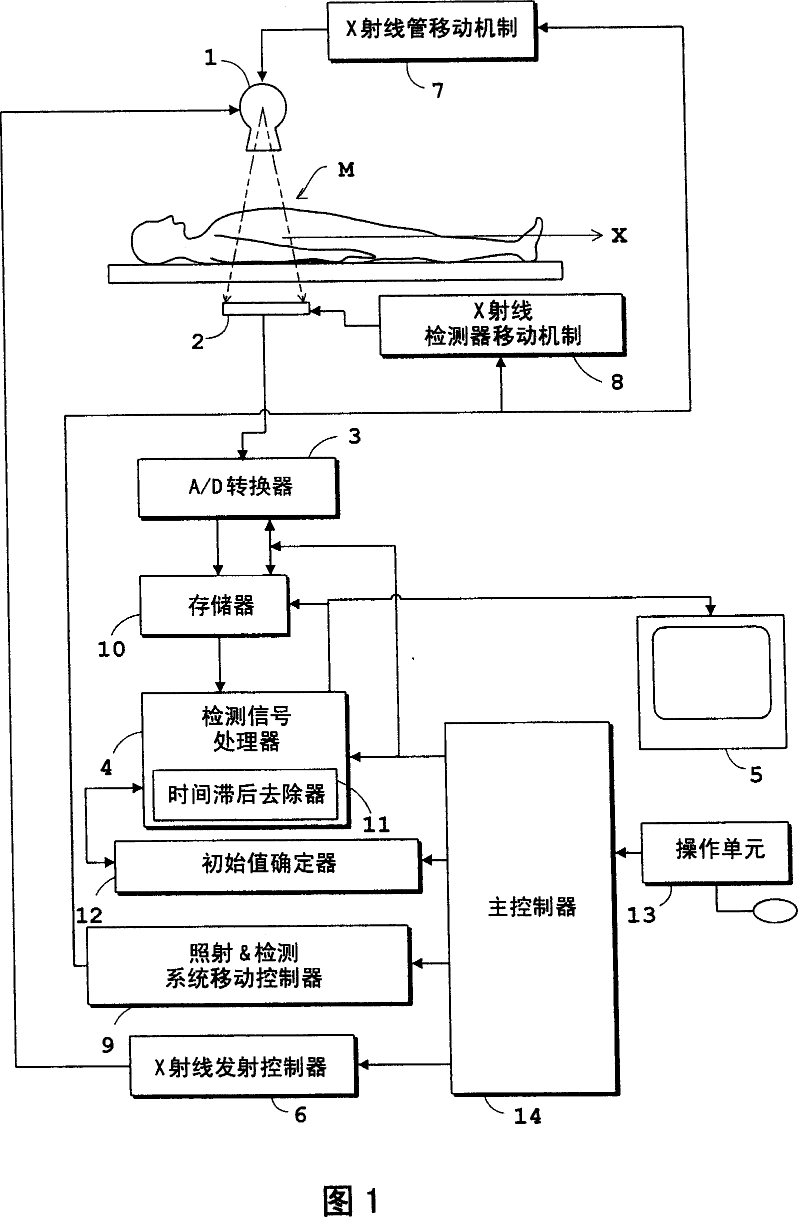
[0205] FIG. 1 is a block diagram showing the overall structure of a fluoroscopy apparatus according to the present invention.

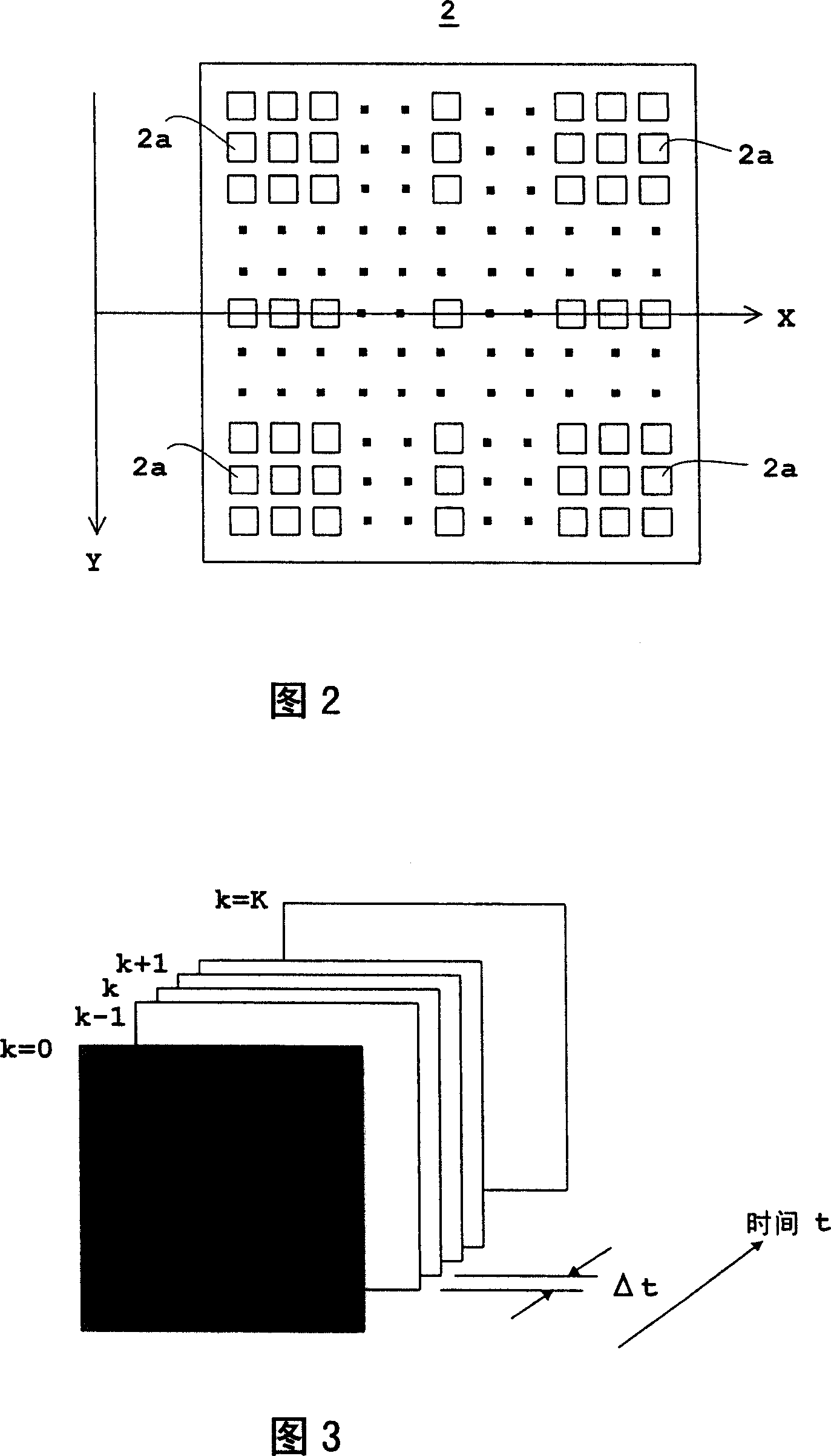
[0206]As shown in FIG. 1 , the fluoroscopy apparatus includes: an X-ray tube 1 for emitting X-rays to a patient M; an FPD (Flat Panel X-ray Detector) 2 for detecting X-rays transmitted through the patient M; an analog-to-digital converter 3, for digitizing the X-ray signal acquired from the FPD2 at a predetermined sampling time interval Δt; a detection signal processor 4, for creating an X-ray image based on the X-ray detection signal output from the analog-to-digital converter 3; and an image monitor 5, for displaying the X-ray image created by the detection signal processor 4. That is, the present apparatus is configured to obtain an X-ray image based on the X-ray detection signal acquired from the FPD 2 by t...


PUM


Abstract

Corrected X-ray detection signals are obtained by removing lag-behind parts through a recursive computation based on initial values determined from lag signal value (step T 2). Thus, the lag-behind parts can be removed by taking into consideration lag signal values remaining at a starting point of the recursive computation. The lag signal values are dependent on the characteristic of an FPD (flat panel X-ray detector). By removing the lag-behind parts, with the lag signal values taken into consideration, the lag-behind parts are removed from X-ray detection signals with increased accuracy, without being influenced by the characteristic of the FPD.

Description

technical field [0001] The present invention relates to a radiographic apparatus for medical or industrial use and a radiation detection signal processing method for outputting radiation at predetermined sampling time intervals from a radiation emitting device when radiation is emitted to a subject. The radiation detection signal obtains a radiographic image. More specifically, the present invention relates to a technique for eliminating a time lag of a radiation detection signal output from the radiation detection device caused by the radiation detection device. Background technique [0002] In recent years, in medical X-ray diagnostic equipment as an example of a typical radiography equipment, a flat panel X-ray detector (hereinafter referred to as "FPD") has been used for detecting X-rays of a patient generated by X-rays emitted from an X-ray tube. X-ray inspection device for radiation penetration images. The FPD includes a plurality of semiconductor or other X-ray dete...

Claims


Application Information

Patent Timeline
Patent Type & Authority Applications(China)
IPC IPC(8): A61B6/00G01N23/04G03B42/00G01T1/161
CPCH04N5/3597H04N5/32A61B6/585H04N5/374H04N25/626H04N25/76H04N23/30G01J1/42A61B6/00
Inventor 冈村升一
Owner SHIMADZU SEISAKUSHO LTD
Why Eureka
Try Eureka