Advanced methods and systems for treating cell proliferation disorders

a cell proliferation and advanced technology, applied in the field of advanced methods and systems for treating cell proliferation disorders, can solve the problems of limiting affecting the effectiveness of current chemotherapy, and being expected to be an even greater threat to our society and economy

Active Publication Date: 2014-07-08
IMMUNOLIGTHT LLC +1
View PDF14 Cites 22 Cited by
  • Summary
  • Abstract
  • Description
  • Claims
  • Application Information

AI Technical Summary

Benefits of technology

[0066]Accordingly, one object of the present invention is to provide a method for the treatment of a cell proliferation disorder that permits treatment of a subject in any area of the body while being non-invasive and having high selectivity for targeted cells relative to healthy cells through the use of plasmonics materials.
[0070]A further object of the present invention is to provide a method for the treatment of a cell proliferation disorder that permits treatment of a subject in any area of the body while being non-invasive and having high selectivity for targeted cells relative to healthy cells through the use of exciton-plasmon enhancement.
[0073]A further object of the present invention is to provide a method for generating an autovaccine effect in a subject, which can be in vivo thus avoiding the need for ex vivo treatment of subject tissues or cells, or can be ex vivo.

Problems solved by technology

It has been one of the principal causes of death in developed countries and, as our population continues to age, it is expected to be an even greater threat to our society and economy.
With cancer, although surgery may sometimes accomplish this goal, the propensity of cancer cells to invade adjacent tissue or to spread to distant sites by microscopic metastasis often limits the effectiveness of this option.
Similarly, the effectiveness of current chemotherapy is often limited by toxicity to other tissues in the body.
Most of these cancer treatment methods, including radiation therapy, are known to cause damage to DNA, which if not repaired during a critical stage in mitosis, the splitting of the cell during cell proliferation, leads to a programmed cell death, i.e. apoptosis.
Further, radiation tends to damage healthy cells, as well as malignant tumor cells.
However, given that only a small percentage of the body's lymphocytes are treated, this seems unlikely to be the only mechanism of action.
However, there are still many limitations to ECP.
It requires establishing peripheral intravenous line or central venous access, which may be difficult to do in certain disease states such as systemic sclerosis or arthritis.
There is also a risk of infection at the venous or central line site, or in the central line catheter.
Further, it requires removing typically several hundred milliliters of whole blood from the patient, hence, the treatment is limited to patients who has sufficiently large initial volume of blood to be withdrawn.
Risk of contracting blood-born pathogen (Hepatitis, HIV, etc.) due to exposure to contaminated operating system is also a concern.
A survey of known treatment methods reveals that these methods tend to face a primary difficulty of differentiating between normal cells and target cells when delivering treatment, often due to the production of singlet oxygen which is known to be non-selective in its attack of cells, as well as the need to perform the processes ex vivo, or through highly invasive procedures, such as surgical procedures in order to reach tissues more than a few centimeters deep within the subject.
However, the use of NRI or UV radiation is known to penetrate tissue to only a depth of a few centimeters.
Also, this patent does not describe initiation energy sources emitting energy other than UV, visible, and near infrared energy; energy upgrading other than within the range corresponding to UV and IR light, and downgrading from high to low energy.
However, each method suffers from the drawback that the treatment is dependent on the production of singlet oxygen to produce the desired effect on the tissue being treated, and is thus largely indiscriminate in affecting both healthy cells and the diseased tissue desired to be treated.
These methods are not used for treating a patient and do not teach using photoactivatable agents in conjunction with plasmonics-active agents and / or modulation agents.
However, the method is not suitable for treating a patient and does not teach any mechanisms for stimulating the photosensitizers, indirectly.
However, the reference fails to disclose any mechanism of photoactivation using energy modulation agent that converts the initiation energy to an energy that activates the activatable pharmaceutical agent and also use of other energy wavebands, e.g., X-rays, gamma-rays, electron beam, microwaves or radio waves.
However, the reference fails to address any specific molecules for use in treating lymphomas or any other cancer.
Fluorescing photosensitizers are said to be preferred, but the reference does not teach how to select a system of fluorescent stimulation or photoactivation using fluorescent photosensitizers.
In addition, the reference teaches that 8-MOP is unsuitable for use as an antiviral, because it damages both cells and viruses.
It is well known that UV activation creates such reactive oxygen species, which are capable of seriously damaging otherwise healthy cells.
Both may lead to inactivating damage of tumor cells, viruses and healthy cells.
However, neither, alone or combined, lead to an auto vaccine effect.
While the complexity of the immune response and cytotoxid effects is fully appreciated by researchers, a therapy that harnesses the system to successfully stimulate an auto vaccine effect against a targeted, malignant cell has been elusive, except for extracorporeal photopheresis for treating lymphoma.
U.S. Pat. No. 6,235,508 teaches that 8-MOP and AMT are unacceptable photosensitizers, because each indiscriminately damages both cells and viruses.
However, while a high yield of singlet oxygen is desirable it is by no means sufficient for a photosensitizer to be clinically useful.
Toxicity can become an issue if high doses of photosensitizer are necessary in order to obtain a complete response to treatment.
The preferential accumulation of PDT drug molecules in cancer cells combined with the localized delivery of light to the tumor, results in the selective destruction of the cancerous lesion.
It is not clear that direct damage to DNA is a major effect; therefore, this may indicate that photoactivation of DNA crosslinking is not stimulated efficiently.
Furthermore, when laser light is administered via external illumination of tissue surfaces, the treatment effect of PDT is confined to a few millimeters (i.e. superficial).
The reason for this superficial limitation is mainly the limited penetration of the visible light used to activate the photosensitizer.
However, even these treatments require significantly invasive techniques to treat the surface of the affected organs.
It is possible that the laser light and small amount of remaining microscopic and minimal gross disease results in too little or highly damaged structures.
It is well recognized that a major problem associated with the existing methods of diagnosis and treatment of cell proliferation disorders is in differentiation of normal cells from target cells.
Such target specificity is difficult to achieve by way of surgery since the strategy there is simply to cut out a large enough portion of the affected area to include all diseased cells and hope that no diseased cells have spread to other distant locations.
With chemotherapy, while some degree of differentiation can be achieved, healthy cells are generally adversely affected by chemo-agents.
These high energy beams ionize the atoms which make up a DNA chain, which in turn leads to cell death.
However, the high doses of radiation needed for such therapies damages healthy cells just as effectively as it does diseased cells.
There is no intrinsic means for a radiation beam to differentiate between a healthy cell from a diseased cell either.
However, this procedure has its drawbacks; it requires drawing blood from the patient, thus requiring cumbersome machinery to perform and may require blood transfusion in order to maintain the volume of blood flow in the machine.
Further, this also limits the size of the patient that can be treated, since the extracorporeal volume is great and too much withdrawal of blood increases the risk of hypovolemic shock.
The method is also limited to treating blood-born cell proliferation related disorders such as lymphoma, and is not capable of treating solid tumors or other types of non-blood related cell proliferation disorders.
A problem encountered in PDT therapy is the inability to treat target areas that are more than a few centimeters beneath the surface of the skin without significant invasive techniques, and the fact that PDT typically operates by generation of sufficient quantities of singlet oxygen to cause cell lysis.
However, singlet oxygen in sufficient concentration will lyse not only target cells, but also healthy cells rather indiscriminately.

Method used


Image

  • Advanced methods and systems for treating cell proliferation disorders
  • Advanced methods and systems for treating cell proliferation disorders
  • Advanced methods and systems for treating cell proliferation disorders

Examples


example 1

Preparation of Silver Nanoparticles

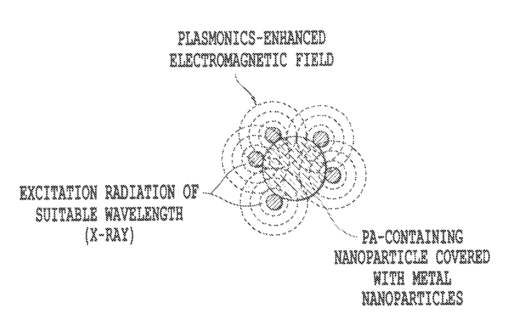
[0498]Silver (or gold) colloids were prepared according to the standard Lee-Meisel method: 200 mL of 10−3 M AgNO3 aqueous solution was boiled under vigorous stirring, then 5 mL of 35-mM sodium citrate solution were added and the resulting mixture was kept boiling for 1 h. This procedure was reported to yield ˜1011 particles / mL of homogenously sized colloidal particles with a diameter of ˜35-50 nm and an absorption maximum at 390 nm. The colloidal solutions were stored at 4° C. and protected from room light. Further dilutions of the colloidal solutions were carried out using distilled water.

Fabrication / Preparation of Metal Nanocaps

[0499]One approach has involved the use of nanospheres spin-coated on a solid support in order to produce and control the desired roughness. The nanostructured support is subsequently covered with a layer of silver that provides the conduction electrons required for the surface plasmon mechanisms. Among the techniques base...

example 2

[0518]Vitamin B12 is used as a stimulating energy source for a photoactive agent overlapping its emission wavelength using dipole-dipole resonance energy transfer.

[0519]

Excitation Max.Emission Max.Endogenous Fluorophore(nm)(nm)Vitamin B12275305

[0520]Vitamin B12 has an excitation maximum at about 275 nm and an emission maximum at 305 nm, as shown above and in Table 2. In this example, 113Sn and / or 137Cs are chelated with the Vitamin B12. The Vitamin B12 preferentially is absorbed by tumor cells. Thus, it is in close proximity and capable of activating 8-MOP, which is administered in advance as the photoactivation molecules. The emission band of Vitamin B12 overlaps the excitation band of 8-MOP; therefore, photo and resonance energy transfer occurs, when Vitamin B12 is in close proximity to 8-MOP. 8-MOP is activated and binds to DNA of the tumor cells inducing an auto vaccine effect in vivo.

example 3

[0521]In this example, gold nanoparticles are chelated with the Vitamin B12 complex. A suitable light source is used to stimulate the gold nanoparticles or Vitamin BI2 may be chelated with one of the UV emitters listed in Table 4 in addition to the gold nanoparticles. The tumor cells preferentially absorb the Vitamin B12 complexes, such that the activated gold nanoparticles are within 50 nanometers of 8-MOP and / or other photoactivatable molecules previously administered. Therefore, resonance energy transfer activates the photoactivatable molecules, such as 8-MOP, and the activated 8-MOP binds to DNA in tumor cells inducing apoptosis and autovaccine effects.

[0522]In a further example, the nanoparticles of gold are clusters of 5 gold atoms encapsulated by poly-amidoamine dendrimers. Thus, the gold nanoparticles emit UV in the correct band for activating 8-MOP and other UV-activatable agents capable of exhibiting photopheresis and / or photodynamic effects.

[0523]Cells undergoing rapid pr...


PUM

PropertyMeasurementUnit
volumeaaaaaaaaaa
wavelengthaaaaaaaaaa

Abstract

The present invention relates to methods for treating cell proliferation disorders comprising (1) administering to the subject at least one activatable pharmaceutical agent that is capable of activation by a simultaneous two photon absorption event and of effecting a predetermined cellular change when activated, (2) administering at least one plasmonics-active agent to the subject, and (3) applying an initiation energy from an initiation energy source to the subject, wherein the plasmonics-active agent enhances or modifies the applied initiation energy, such that the enhanced or modified initiation energy activates the activatable pharmaceutical agent by the simultaneous two photon absorption event in situ, thus causing the predetermined cellular change to occur, wherein said predetermined cellular change treats the cell proliferation related disorder, and the use of plasmonics enhanced photospectral therapy (PEPST) and exiton-plasmon enhanced phototherapy (EPEP) in the treatment of various cell proliferation disorders, and the PEPST and EPEP agents and probes.

Description

CROSS REFERENCE TO RELATED APPLICATIONS[0001]This application claims priority to Provisional Application Ser. No. 61 / 080,429 filed Jul. 14, 2008 the content of which is hereby incorporated herein by reference. U.S. application Ser. No. 11 / 935,655, filed Nov. 6, 2007; and Provisional Application Ser. No. 60 / 954,263, filed Aug. 6, 2007, and 61 / 030,437, filed Feb. 21, 2008, 61 / 042,561, filed Apr. 4, 2008; 61 / 035,559, filed Mar. 11, 2008, and 61 / 080,140, filed Jul. 11, 2008, and U.S. application Ser. No. 12 / 059,484, filed Mar. 31, 2008, are related to this application and are each hereby incorporated by reference.BACKGROUND OF THE INVENTION[0002]1. Field of Invention[0003]The present invention relates to methods and systems for treating cell proliferation disorders, that provide better distinction between normal, healthy cells and those cells suffering a cell proliferation disorder (hereafter “target cells”) and preferably that can be performed using non-invasive or minimally invasive t...

Claims


Application Information

Patent Timeline
Patent Type & Authority Patents(United States)
IPC IPC(8): A61N5/10A61N5/06A61N5/00
CPCA61K31/7056A61N5/062A61K47/48107A61K41/008A61K47/551A61K31/4427A61K31/7052A61P35/00A61K47/55
Inventor BOURKE, JR., FREDERIC A.VO DINH, TUAN
Owner IMMUNOLIGTHT LLC
Why Eureka
Try Eureka