Endoscope system and low visibility determining method
a technology of endoscope and determining method, which is applied in the field of endoscope system and low visibility determining method, can solve problems such as smoke or mist generation
- Summary
- Abstract
- Description
- Claims
- Application Information
AI Technical Summary
Benefits of technology
Problems solved by technology
Method used
Image
Examples
first embodiment
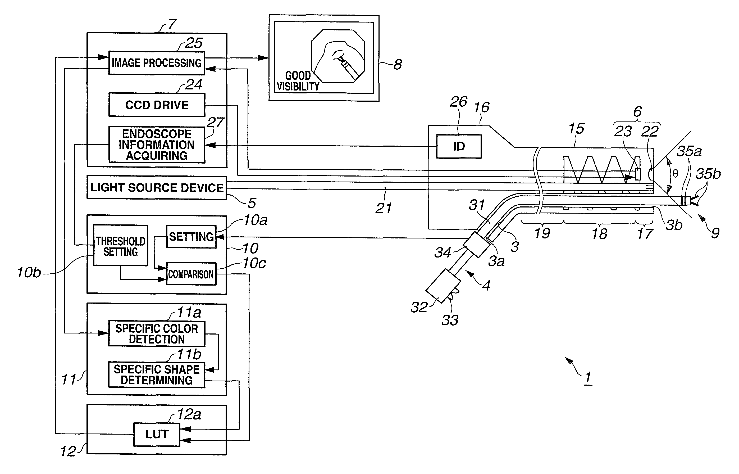
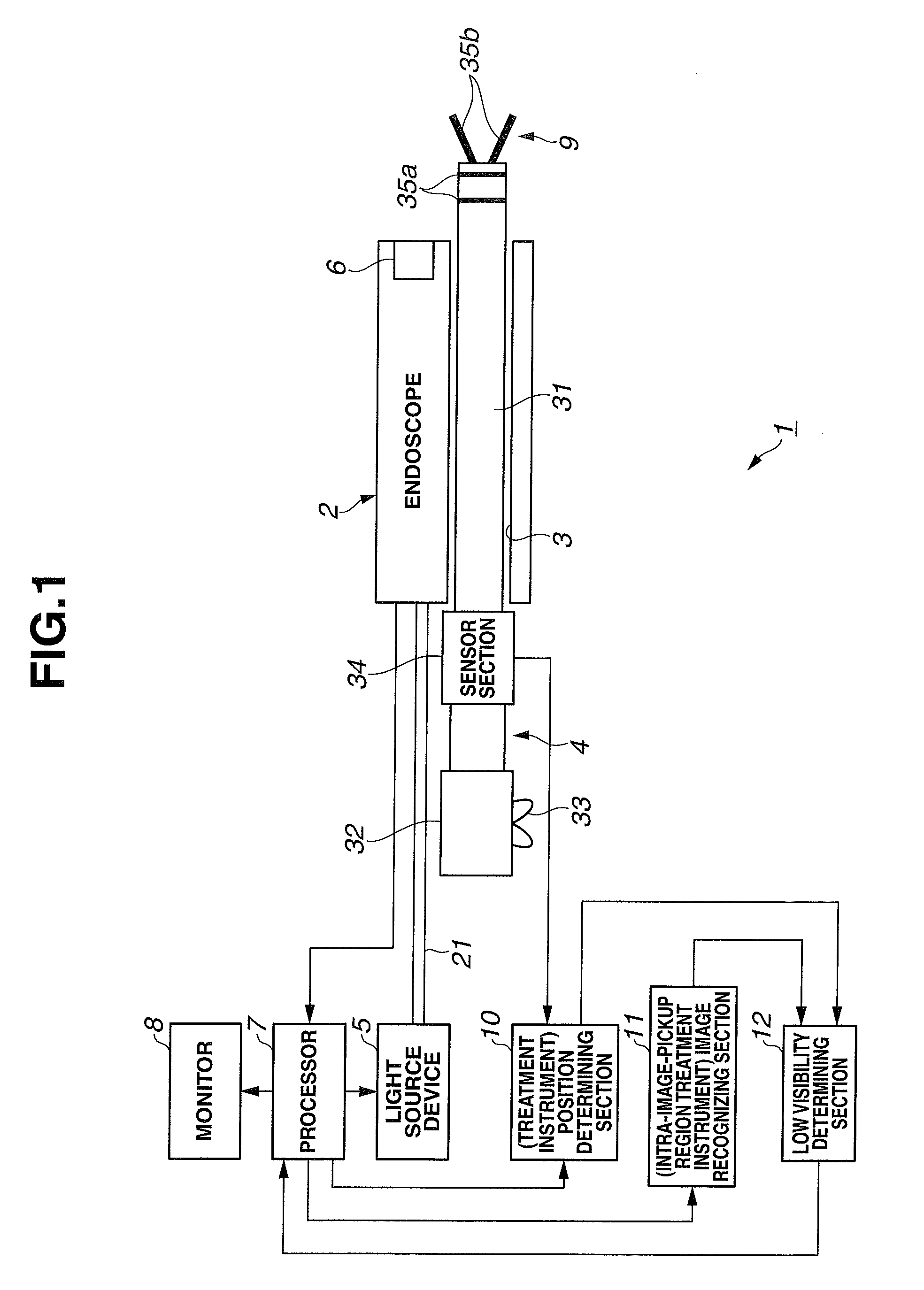
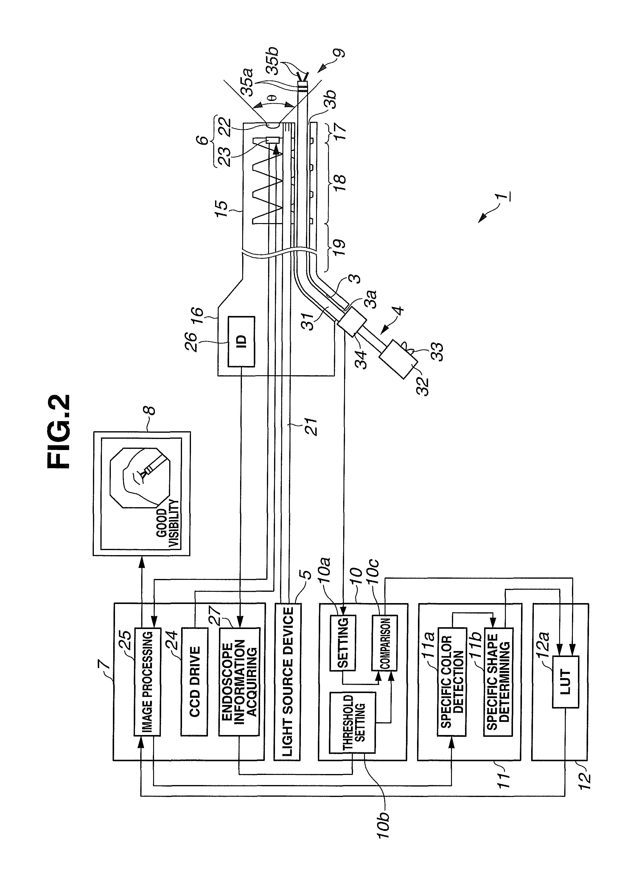
[0032]As shown in FIG. 1, an endoscope system 1 according to a first embodiment of the present invention includes an endoscope 2 inserted into a body, a treatment instrument 4 inserted into a treatment instrument channel (simply abbreviated as “channel”) 3 of the endoscope 2 and a light source device 5 that supplies illuminating light to the endoscope 2.
[0033]Furthermore, this endoscope system 1 includes a processor 7 as signal processing means for performing signal processing with respect to an image pickup section 6 as image pickup means provided in the endoscope 2 and a monitor 8 as display means for displaying an endoscope image corresponding to an image signal generated by the processor 7.
[0034]The light source device 5 and the processor 7 may be connected together via a signal line and a light quantity of illuminating light from the light source device 5 may be adjusted by a brightness signal of the image signal from the processor 7.
[0035]Furthermore, this endoscope system 1 i...
second embodiment
[0096]Next, a second embodiment of the present invention will be described with reference to FIG. 5 to FIG. 9. FIG. 5 illustrates an overall configuration of an endoscope system 1B according to the second embodiment of the present invention.
[0097]The endoscope system 1B shown in FIG. 5 corresponds to the endoscope system 1 in FIG. 1 using a high-frequency treatment instrument 4B that performs a treatment using high-frequency electric energy instead of the treatment instrument 4 and this high-frequency treatment instrument 4B is connected to a high-frequency power supply apparatus 41 as an electric energy supply apparatus.
[0098]Furthermore, this high-frequency power supply apparatus 41 is connected to a foot switch (abbreviated as “FS” in FIG. 5) 42 that performs an instruction operation to output or stop outputting a high-frequency current (ON / OFF). The high-frequency treatment instrument 4B, the high-frequency power supply apparatus 41 and the foot switch 42 constitute a high-frequ...
third embodiment
[0158]Next, a third embodiment of the present invention will be described with reference to FIG. 10 to FIG. 12. FIG. 10 is a diagram illustrating an overall configuration of an endoscope system 1C according to the third embodiment of the present invention.
[0159]The endoscope system 1C corresponds to the endoscope system 1B according to the second embodiment shown in FIG. 5 using an active high-frequency treatment instrument (simply abbreviated as “high-frequency treatment instrument”) 4C provided with a drive section 50 that actively drives a moving section instead of the manual high-frequency treatment instrument 4B. This high-frequency treatment instrument 4C, a high-frequency power supply apparatus 41 and a foot switch 42 form a high-frequency electrocoagulation apparatus 43C.
[0160]Furthermore, the endoscope system 1C of the present embodiment is provided with a control section 51 that controls operation of the high-frequency treatment instrument 4C and also controls an energy su...
PUM
 Abstract
Description
Claims
Application Information
 - R&D
- Intellectual Property
- Life Sciences
- Materials
- Tech Scout
- Unparalleled Data Quality
- Higher Quality Content
- 60% Fewer Hallucinations
Browse by: Latest US Patents, China's latest patents, Technical Efficacy Thesaurus, Application Domain, Technology Topic, Popular Technical Reports.
© 2025 PatSnap. All rights reserved.Legal|Privacy policy|Modern Slavery Act Transparency Statement|Sitemap|About US| Contact US: help@patsnap.com


