
Method and apparatus for levator distension repair

a technology of levator and distension, applied in the field of urogenital surgery, can solve the problems of “dragging feeling”, increased importance of problems, and long-standing female genital prolaps

Inactive Publication Date: 2012-09-11
BOSTON SCI SCIMED INC
View PDF51 Cites 10 Cited by
  • Summary
  • Abstract
  • Description
  • Claims
  • Application Information

AI Technical Summary

Benefits of technology

The present invention is about surgical instruments and implantable articles for urological applications, particularly for repairing pelvic organ prolapse. The invention includes a method of placing mesh subcutaneously against the levator muscle, which is a new and improved technique. The mesh is placed without needing to dissect the vagina. The mesh is shaped like a trapezoid and is secured with sutures or staples after being implanted subcutaneously. The technical effects of this invention include improved outcomes, reduced complications, and reduced recovery time for patients.

Problems solved by technology

Female genital prolapse has long plagued women.
With the increasing age of the U.S. population, these problems will likely assume additional importance.
The common clinical symptoms of vaginal prolapse are related to the fact that, following hysterectomy, the vagina is inappropriately serving the role of a structural layer between intra-abdominal pressure and atmospheric pressure.
This pressure differential puts tension on the supporting structures of the vagina, causing a “dragging feeling” where the tissues connect to the pelvic wall or a sacral backache due to traction on the uterosacral ligaments.
Exposure of the moist vaginal walls leads to a feeling of perineal wetness and can lead to ulceration of the exposed vaginal wall.
Vaginal prolapse may also result in loss of urethral support due to displacement of the normal structural relationship, resulting in stress urinary incontinence.
Certain disruptions of the normal structural relationships can result in urinary retention, as well.
Stretching of the bladder base is associated with vaginal prolapse and can result in complaints of increased urinary urgency and frequency.
Anterior vaginal wall prolapse causes the vaginal wall to fail to hold the bladder in place.
There may also be a transverse defect, causing cystocele across the vagina.
They may occur because of failure to reapproximate the superior aspects of the pubocervical fascia and the rectovaginal fascia at the time of surgery.
Mid-vaginal and high rectoceles may result from loss of lateral supports or defects in the rectovaginal septum.
Further, loss of connective tissue strength might be associated with damage at childbirth, deterioration with age, poor collagen repair mechanisms, and poor nutrition.
As noted, vaginal prolapse and the concomitant anterior cystocele can lead to discomfort, urinary incontinence, and incomplete emptying of the bladder.
Posterior vaginal prolapse may additionally cause defecatory problems, such as tenesmus and constipation.
Furthermore, apart from the physical symptoms, vaginal prolapse has been shown to result in a lower quality of life for its sufferers, including feeling less attractive, less feminine, and less sexually attractive.
Increases in abdominal pressure, failure of the muscles to keep the pelvic floor closed, and damage to the ligaments and fascia all contribute to the development of prolapse.
In addition, if a woman has a hysterectomy, the vaginal angle may be altered, causing increased pressure at a more acute angle, accelerating the prolapse.
Damage to the cardinal uterosacral ligament complex permits the uterus and upper vagina to telescope downwards, like an inverted sock.
Complete failure of the cardinal uterosacral ligament complex will result in a “cervix-first” prolapse.
Traditional anterior prolapse repairs have a relatively high failure rate.
Such transvaginal dissection can be more difficult for the surgeon and may lead to further failures.

Method used


Image

  • Method and apparatus for levator distension repair
  • Method and apparatus for levator distension repair
  • Method and apparatus for levator distension repair

Examples


Embodiment Construction

[0036]Referring now to the drawings, wherein like reference numerals designate identical or corresponding parts throughout the several views. The following description is meant to be illustrative only, and not limiting other embodiments of this invention will be apparent to those of ordinary skill in the art in view of this description.

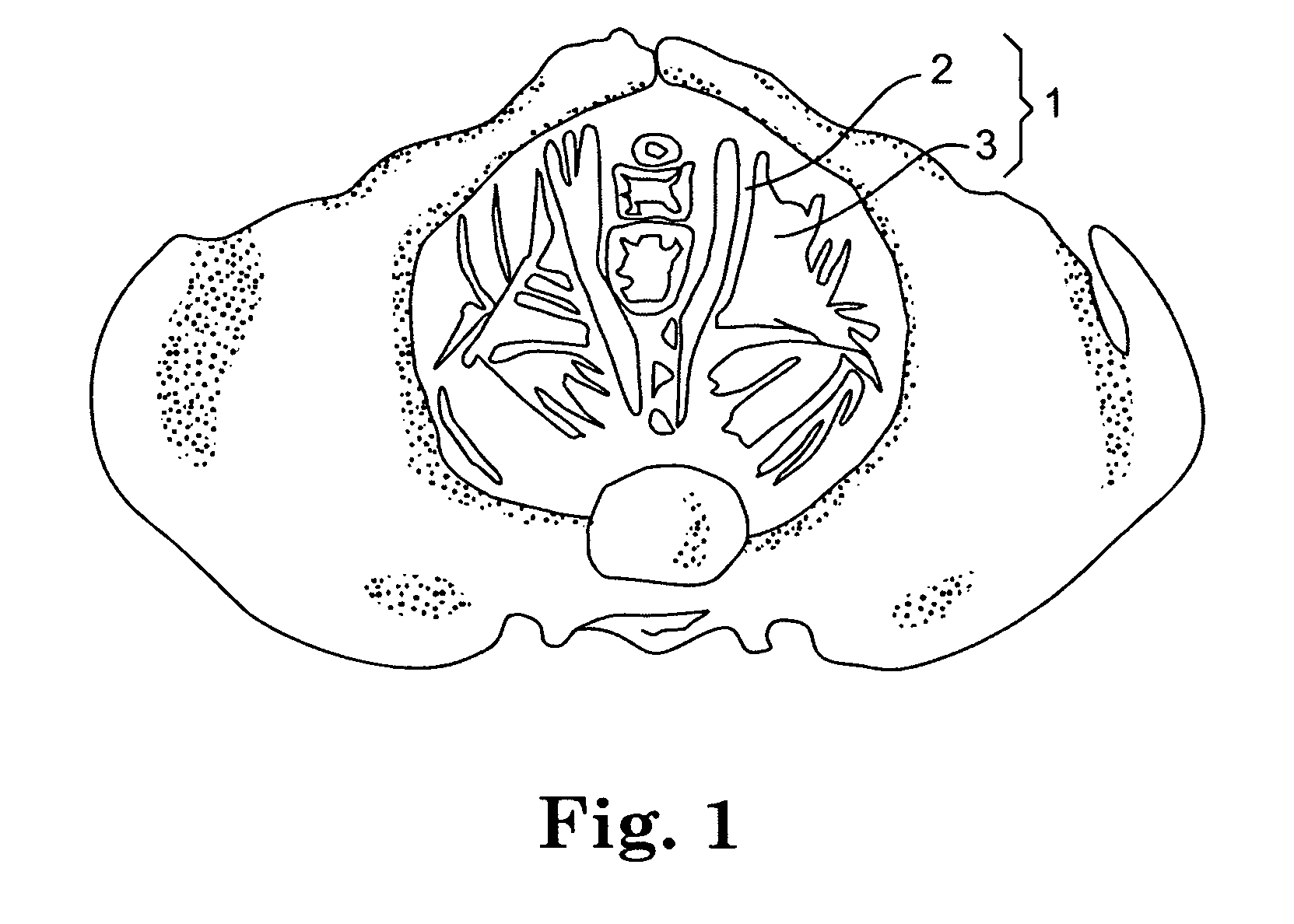
[0037]The relevant anatomy is illustrated in FIG. 1. As can be seen, the levator ani muscles 1, including the pubococcygeus 2 and illiococcygeus muscles 3, are a significant portion of the pelvic floor and provide support for the pelvic viscera. FIGS. 1 and 2 show the normal condition of the levator muscles, while FIG. 3 shows the posture of levator muscles associated with prolapsed pelvic organs. As can be seen, such muscles offer less support for the pelvic viscera and may benefit from additional support as provided in the present invention.

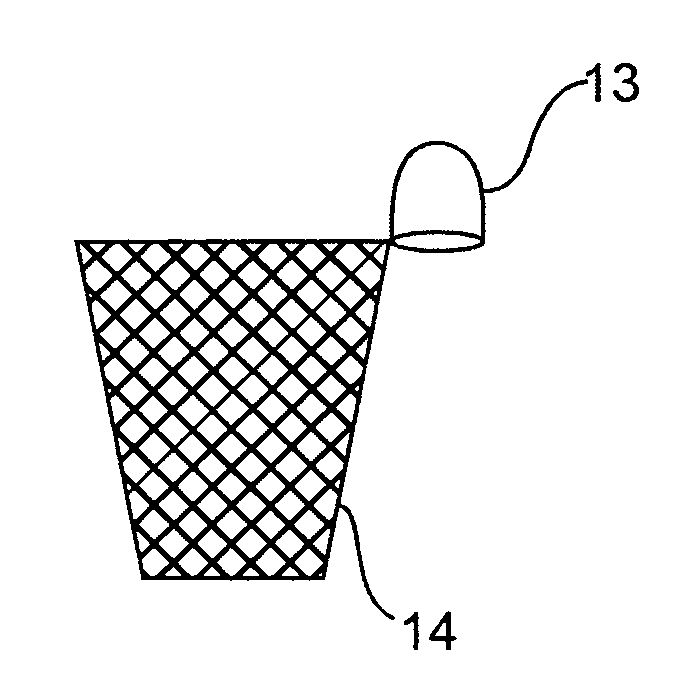
[0038]In the present invention, the mesh implant, an embodiment which is illustrated in FIG. 4, is placed exte...


PUM


Abstract

Improved methods and apparatuses for treatment of pelvic organ prolapse are provided. A specialized mesh having a shape for convenient subcutaneous placement to support the levator ani muscles is provided, as is a method of use of such a device. Appropriate devices for introducing such a mesh implant are also disclosed.

Description

PRIORITY[0001]This application claims benefit from International Application No. PCT / US2007 / 070732, having PCT Publication No. WO 2007 / 146784, which was filed on 8 Jun. 2007, which in turn claims priority under 35 USC §119(e) to U.S. Provisional Application Ser. No. 60 / 811,790, filed 8 Jun. 2006 and to U.S. Provisional Application Ser. No. 60 / 825,357, filed 12 Sep. 2006, the entire content of each application being incorporated herein by reference.BACKGROUND OF THE INVENTION[0002]1. Field of the Invention[0003]This invention relates to urogenital surgery.[0004]2. Description of the Related Art[0005]Female genital prolapse has long plagued women. It is estimated by the U.S. National Center for Health Statistics that 247,000 operations for genital prolapse were performed in 1998. With the increasing age of the U.S. population, these problems will likely assume additional importance.[0006]The common clinical symptoms of vaginal prolapse are related to the fact that, following hysterect...

Claims


Application Information

Patent Timeline
Patent Type & Authority Patents(United States)
IPC IPC(8): A61F2/00
CPCA61B17/0401A61F2/0045A61B2017/00438A61B2017/00805A61B2017/0408
Inventor CHAPMAN, KELLY ANNOLSON, MATTHEW J.HALLUM, III, ALTON V.
Owner BOSTON SCI SCIMED INC
Learn More