Method for processing biomedical images

a biomedical image and image processing technology, applied in image data processing, character and pattern recognition, instruments, etc., can solve the problems of increasing tumour incidence, difficult early diagnosis of tumours, and real social problems, and achieve the effect of reducing management costs

Inactive Publication Date: 2012-03-27
MEDICAD
View PDF3 Cites 4 Cited by
  • Summary
  • Abstract
  • Description
  • Claims
  • Application Information

AI Technical Summary

Benefits of technology

[0011]For this purpose, one performs the early diagnosis through the so-called “screening programs” which consist in applying diagnostic tests to the asymptomatic patients, with the purpose to identify the subjects potentially inclined to develop the disease. The ultimate screening objective is to reduce the specific mortality of the investigated pathology among the population making the object of study, thanks to early diagnosis and to neoplasm treatment in its initial phase.
[0023]to improve the mammographic sensitivity, through the increasing of the meaningful potentially suspicious areas detection;
[0024]to improve the specificity in order to decrease the number of False Positives, thus reducing the number of unnecessary biopsies.
[0032]Images segmentation is a key problem in “computer vision”. Such action allows the splitting of the image in regions corresponding to objects or parts of objects represented in it, and it acts as a first step towards the extraction of meaningful information. The segmentation is a decomposition of the image in spatially separated components. The pixels in each component exhibit some similarities of their characteristics, as for instance their grey levels. The two main image segmentation techniques are based on the contour or on the region. In the contour based approach, the information is found by the detection of the boundary. On the other hand, the main idea of region segmentation is the identification inside the image of various regions with similar characteristics. The initial segmentation will strongly influence the following phases of the classification process. Therefore such segmentation has to be the most accurate possible, avoiding both the under-segmentation (merging in the same zone of semantically different objects) and the over-segmentation (splitting of a semantically unique object in different zones).
[0042]The method described in this invention is set as a tool for the early diagnosis of mass lesions in mammography. The invention can be used in the context of a CAD that operates as second reader in the normal clinical routine and in the mammographic screening, reducing the costs of management for the “double reading” procedure.

Problems solved by technology

The current way of life, the atmospheric pollution, the chemical additives used for food conservation and other factors are surely the main causes of the enormous increase of tumour incidence during the last 50 years: one may state that the tumours became a real social problem.
At the beginning, the breast cancer appears as a single and confined lesion (usually called “early” or “minimal” breast cancer) and its dimensions do not exceed one centimeter; the lack of certain symptomatic signals of the disease makes difficult its early diagnosis.
Nevertheless the methodology, although notably improved over the years, cannot ensure recognition of all pathologic breast lesions: more recent statistics report percentages varying between 10% and 30% of tumours not being diagnosed with the mammographic examination.
The identification of such lesions raises several problems related to intrinsic characteristics of the lesions:dimensions, forms and density may vary on a high scale range;very often the boundaries are not well defined;they show up high likeness to the parenchyma structure.
On the other hand, the variety and the complexity of such connective and glandular structures make the extraction of the signal an arduous task for the radiologist.
Some structural problems put a limit to the potentialities of the two latter families of algorithms.
In fact, for the type “2” algorithms, in the case of dense tissue one gets very often a higher number of suspicious regions, with a consequent increase of the false positives per image.
As for the type “3” algorithms, very often the two breasts are not perfectly super-imposable because of their intrinsic differences.
Images segmentation is a key problem in “computer vision”.

Method used


Image

  • Method for processing biomedical images
  • Method for processing biomedical images
  • Method for processing biomedical images

Examples


Embodiment Construction

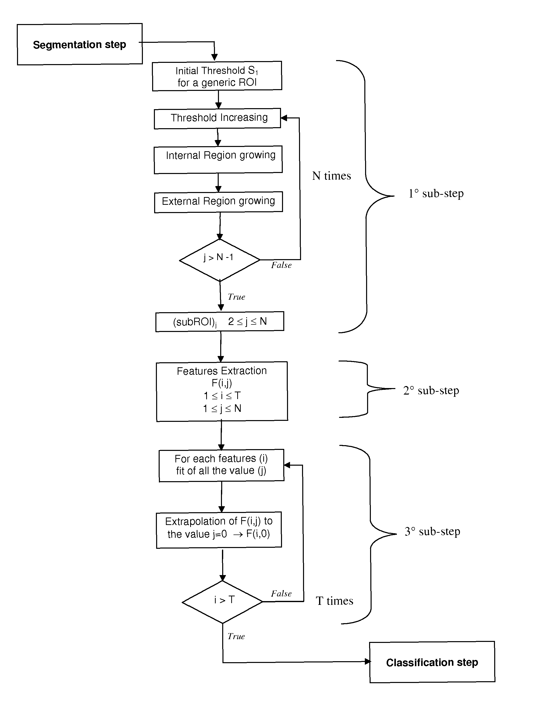
[0055]Generally speaking, the functioning of a CAD can be divided in different steps that can be schematized as shown in FIG. 2, in which is represented a flow diagram of the method according to the present invention. The steps are the folio wings:[0056]pre-processing: the digital image is processed in order to delimit the mammographic area to be submitted to further analysis;[0057]segmentation: the cleaned image is segmented in order to select regions of interest (ROI) mapping out its contour;[0058]extraction: from each ROI is extracted some characteristic information;[0059]classification: to each ROI is assigned a probability of pathology;[0060]visualization: the full mammographic image is displayed on a screen or on paper, highlighting the ROIs having a pathology probability superior to a threshold value selected by the radiologist.

[0061]According to this invention, the extraction step is divided in 3 more sub-steps, as it will be explained in details:[0062]1° sub-step: N subROIj...


PUM


Abstract

The present invention relates to a method for highlight and to diagnose regions of interest in biomedical radiographic images, useful in the context of a CAD tool processing operating as second reader during the normal clinical and screening routine, so reducing the costs of management of the “double reading” procedure.

Description

CROSS REFERENCE TO RELATED APPLICATIONS[0001]The present application is the US national stage of International Application PCT / IB2007 / 051282 filed on Apr. 10, 2007 which, in turn, claims priority to Italian Application RM2006A000213, filed on Apr. 13, 2006.INTRODUCTION[0002]The present invention applies to a processing method of biomedical images for the early detection of tumoral pathologies, which is included in a CAD for diagnostic aid.[0003]The further described invention can be used, for instance, to look for mass lesions in mammographic images, but it can be used also to look for other generic mammary pathologies and tumoral forms.[0004]Moreover, the further described methodology is enough flexible to adapt it too to the analysis of other typical medical diagnostic images: generic radiographies, spiral CT, NMR, ecographies, etc.[0005]From the Greek καρκινοσ (crab), the word carcinoma is owed to Hippocrates and designs the infiltration characteristic of a tumour and its extraor...

Claims


Application Information

Patent Timeline
Patent Type & Authority Patents(United States)
IPC IPC(8): G06K9/00G06K9/32
CPCG06T7/0012G06T7/0081G06T2207/10116G06T2207/30068G06T2207/30196G06T7/11
Inventor CASCIO, DONATOFAUCI, FRANCESCOMAGRO, ROSARIORASO, GIUSEPPE
Owner MEDICAD
Why Eureka
Try Eureka