
Method for image processing and contour assessment of the heart

a heart and contour assessment technology, applied in the field of image processing and contour assessment of the heart, can solve the problems of increasing morbidity of patients, increasing the risk of complications,

Inactive Publication Date: 2010-04-06
CHASE MEDICAL +1
View PDF134 Cites 220 Cited by
  • Summary
  • Abstract
  • Description
  • Claims
  • Application Information

AI Technical Summary

Benefits of technology

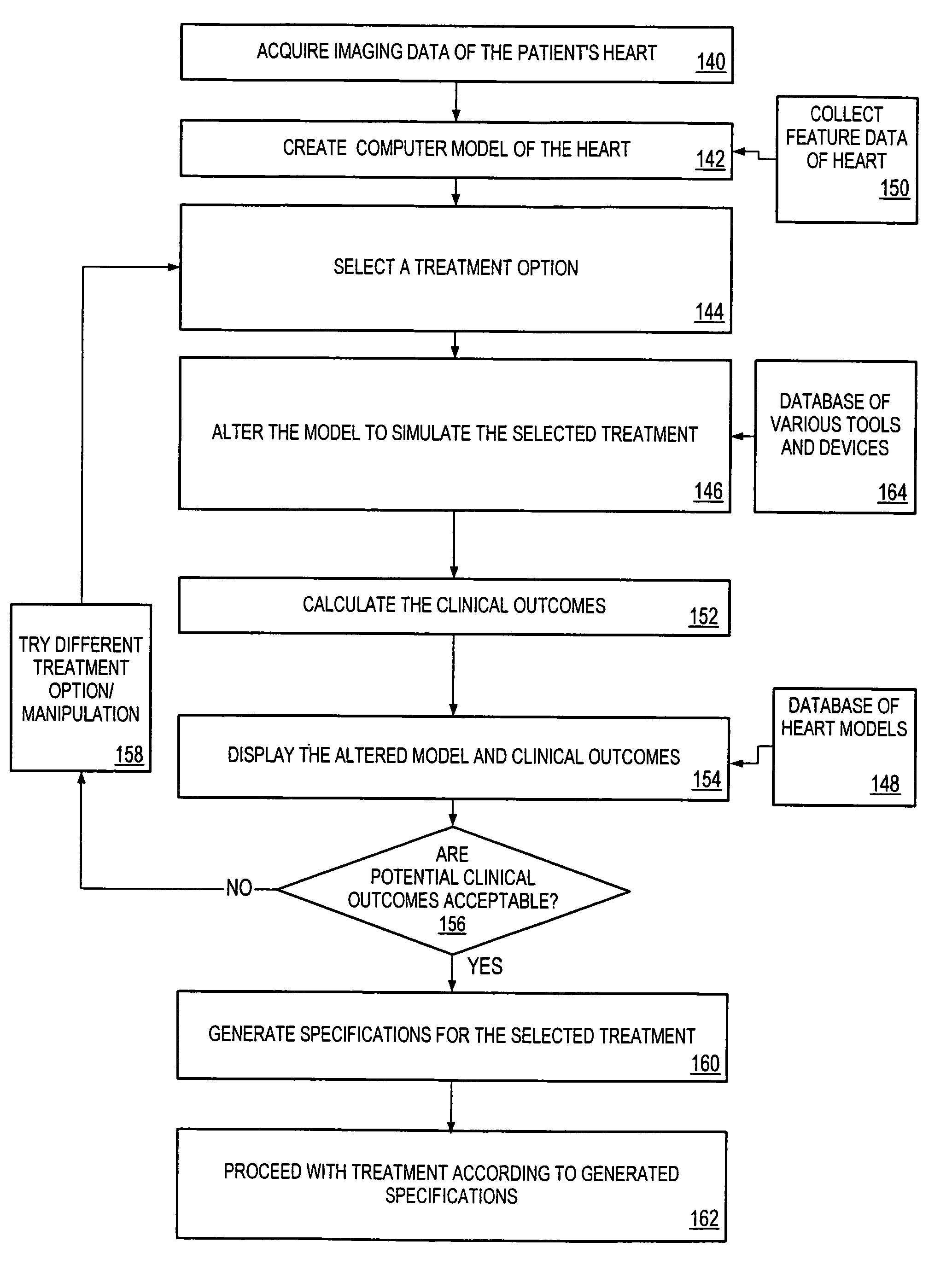
[0019]In an embodiment, a method of designing cardiac instruments may include providing at least one image of heart tissue from a human heart to a computer system. A method may include creating a pattern of at least a portion of at least one cardiac instrument using at least one image. In some embodiments, a plurality of images may be included and an at least three-dimensional model created from some of the images. An image of the results may be created by a computer system. Images created by a computer system of a design of a cardiac instrument may be used to assist in the manufacturing of the instrument.

Problems solved by technology

This increases morbidity to the patient and may contribute to death of the patient who may not tolerate the additional stress of a longer, more invasive procedure.
On some patients, the extra grafts may be needed, since collateral flow, or flow from other arteries, is not sufficient to perfuse the entire myocardium.
Currently, the user has no way. of knowing if the total number of grafts that he put in was appropriate.
There is no precise way to determine how much a valve or structural component needs to change or what the effect of that change will be.
This procedure subjects the patient to unnecessary risks by exposing them to longer than necessary bypass runs and reperfusion injuries each time the heart is weaned of cardioplegia.
This procedure also takes up valuable operating room and staff time.
Additionally, this assessment method only assesses one factor related to the performance of the valve and ventricle regurgitation.
A user may perform a procedure, which corrects the existing problem, but the procedure may create another problem or diminish the performance of the ventricle.
The user has little, if any, way to know if he compromised ventricle performance, since current analytical tools only look for flow across the valve.

Method used


Image

  • Method for image processing and contour assessment of the heart
  • Method for image processing and contour assessment of the heart
  • Method for image processing and contour assessment of the heart

Examples


caselles

[0453 has shown that parametric active contours and geometric contours are basically equivalent and that one formulation can be derived from the other. One basic difference, however, is in the handling of what is sometimes called “the topology problem.” In particular, when there are several objects in the scene, the topology of the final curve is object-dependent and the algorithm must adjust. In the geometric active contour formulation, this is handled automatically since the parameterization of the level-set is calculated only after convergence. In the parametric active contour formulation, more extensive measures need to be taken to split active contours into two or more pieces or to instantiate separate active contours at different locations. Except for topological differences, however, the behavior of both active contour models is fundamentally characterized by the external and internal forces of the formulation.

[0454]There are three key difficulties with active contour algorit...


PUM


Abstract

One embodiment discloses a computerized method of facilitating cardiac intervention. The method may include inputting patient data and creating a computerized interactive model of a heart based on the patient data. The model may include features. The model may simulate at least one proposed cardiac intervention by adding or deleting features to the model, and determining the effects of the proposed cardiac simulation upon the entire model. Simulations may be repeated to allow the user to determine an optimal cardiac intervention. A template and / or patient specific instrument may be created from the model to use as a guide during the cardiac intervention.

Description

PRIORITY CLAIM[0001]This application claims priority to U.S. application Ser. No. 10 / 713,911 entitled “SYSTEM AND METHOD FOR FACILITATING CARDIAC INTERVENTION” filed Jan. 30, 2003 and to U.S. Provisional Patent Application No. 60 / 526,023 entitled “METHOD AND SYSTEM FOR IMAGE PROCESSING AND CONTOUR ASSESSMENT” filed Dec. 1, 2003.RELATED PATENTS[0002]This patent application incorporates by reference in its entirety U.S. patent application Ser. No. 10 / 135,465 entitled “A System and Method for Facilitating Cardiac Intervention,” filed on Apr. 30, 2002.BACKGROUND OF THE INVENTION[0003]1. Field of the Invention[0004]This invention relates generally to systems for identifying the features that contribute to the cardiac performance of an individual patient through the use of imaging methods, and in particular to a computerized system and method for facilitating and assessing cardiac intervention methods.[0005]2. Description of the Related Art[0006]The circulatory system of a human works as ...

Claims


Application Information

Patent Timeline
IPC IPC(8): A61B5/05A61N1/08G06K9/00G06TG06T5/00G06T7/00G16H20/40G16H30/20G16H30/40G16H50/30G16Z99/00
CPCG06F19/3437G06T7/0012G06T7/0083G06F19/3481A61N1/08G06F19/3443G06T2207/30048G06T2207/10088G06T7/12G16H50/50G16H50/70G16H50/30G16H30/20G16H30/40G16H20/40G16Z99/00A61B5/05
Inventor SURESH, MITTADALTON, JUDE
Owner CHASE MEDICAL
Learn More