
Automated high volume slide processing system

a processing system and high-volume technology, applied in chemical methods analysis, applications, instruments, etc., can solve the problems of difficult comparison between different samples, inability of histologists or other medical personnel to interpret slides, and many tissues that do not retain enough color after processing, so as to minimize the possibility of cell carryover and high throughput staining

Active Publication Date: 2008-12-23
VENTANA MEDICAL SYST INC
View PDF294 Cites 201 Cited by
  • Summary
  • Abstract
  • Description
  • Claims
  • Application Information

AI Technical Summary

Benefits of technology

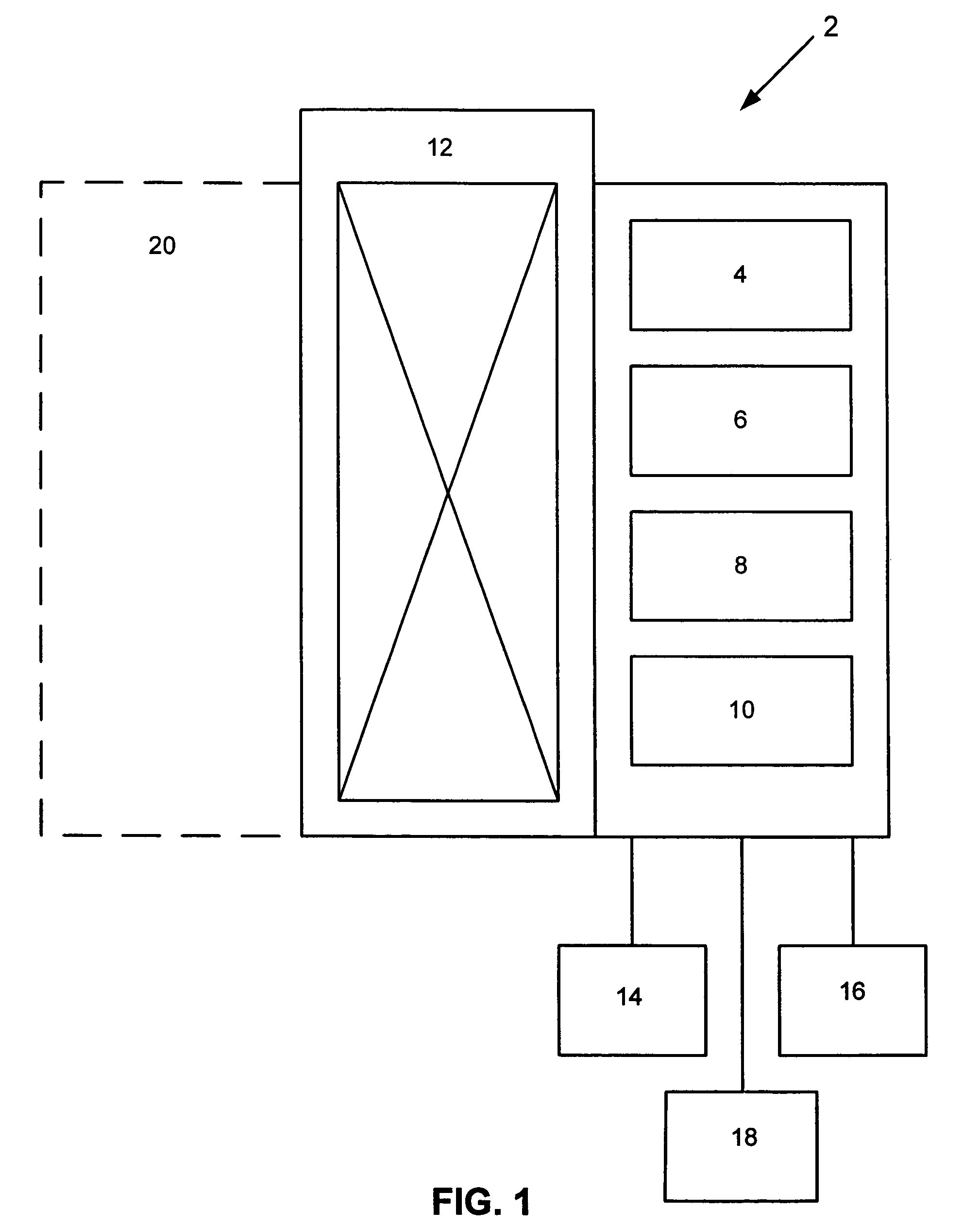
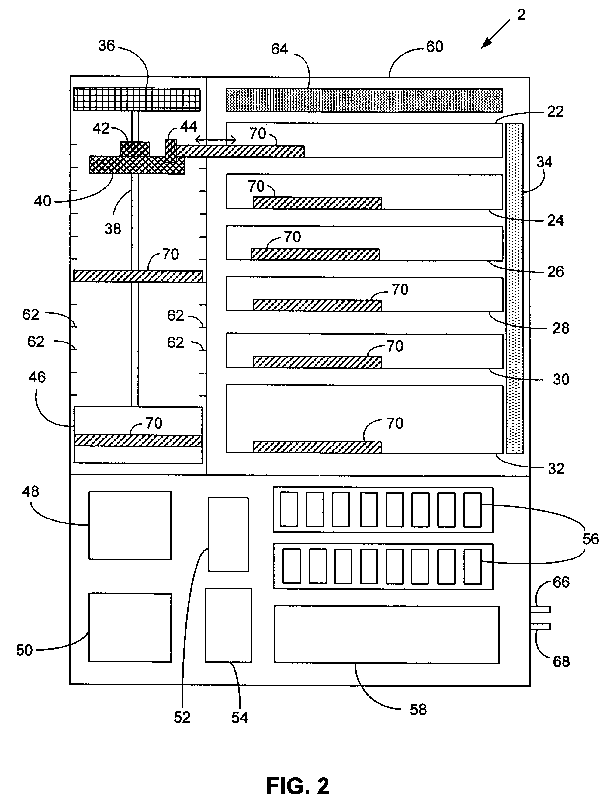
[0010]An automated system is provided for performing slide processing operations on slides bearing biological samples. The system enables high sample throughput and increased staining consistency while also minimizing the potential for cross-contamination of slides.
[0011]In one aspect of the disclosed system, a workstation for performing a step of a staining protocol is not a bath containing a reagent in which several slides are simultaneously immersed. Rather, according to this aspect, a workstation of the system dispenses a reagent to a plurality of microscope slides with minimal transfer of reagent (and contaminants therein) between individual slides. Thus, a workstation according to this aspect minimizes or substantially eliminates the type of cross-contamination of slides that occurs in prior art “dip and dunk” type automated slide staining systems, where contaminants such as dislodged cells can be transferred through the reagent bath from one slide to another.
[0013]In a more particular embodiment, slides are held in a rectangular slide tray in two rows such that their long dimensions are disposed outward from the central, long axis of the tray toward the long edges of the tray. A reagent dispenser in a workstation is positioned above one or more pairs of slides in the opposite rows, and delivers a reagent to one or more slides in one or the other of the two rows, for example, to a pair of slides that are opposite from each other in the two rows. If the reagent dispenser is positioned above fewer than the total number of slides that are held in the tray, the reagent dispenser can move to dispense reagent to other slides in each row of slides, and / or the slide tray can be moved to bring additional slides into position for reagent dispensing. Alternatively, two or more stationary or moving reagent dispensers can be included in the workstation, or one or more manifolds of dispense nozzles can be positioned above the two rows of slides, for example, along the central, long axis of the tray. Nozzles of a reagent dispenser can direct reagent downward and / or upward toward surfaces of slides.
[0017]In another aspect, a fluidics module is disclosed for automated handling of reagents that can deliver reagents in packaged concentration or in diluted concentration to a workstation without the need to disrupt the delivery of such reagents by the workstation while replacing or replenishing reagents to the system. In more particular embodiments, the fluid-handling module includes a dual chamber fluid pump. The dual chamber fluid pump includes a pump chamber and a dispense chamber where the pump chamber is configured to alternate between vacuum and pressure. The two chambers and a set of valves allow the dispense chamber to be maintained at a constant pressure for dispensation of a reagent to slides even while additional reagent is added to the dispense chamber from the pump chamber. Alternatively, a pump chamber supplying a dispense chamber can further function as a dilution chamber, and a concentrate pump chamber can be added to provide concentrated solutions to the dilution chamber.
[0018]The disclosed system is capable of high throughput staining of biological samples on slides without the shortcomings of conventional dip and dunk systems, particularly by eliminating conventional dip-and-dunking de-paraffinizing and / or staining baths, which tend to degrade through oxidation and / or contamination by biological cells dislodged during the de-paraffinizing process. Instead, the disclosed system can employ fresh, clean reagents, thus minimizing the possibility of cell carryover from slide to slide. Moreover, the disclosed system provides for the first time a fully integrated high throughput system for staining slides from the baking step through the coverslipping step, a process that is not performed by any other commercially available system to date.

Problems solved by technology

Many tissues do not retain enough color after processing to make their components visible under a bright-field microscope.
In the past, the steps of staining a tissue sample for histological or cytological analysis were performed manually, a process that is inherently inconsistent.
Inconsistent staining makes it difficult for a Histologist or other medical personnel to interpret slides and to make comparisons between different samples.
A common shortcoming of the automated “dip and dunk” staining devices is the possibility for cross-contamination of samples that are simultaneously or sequentially introduced into the same solution baths.
Another problem inherent to these designs is that as slide baskets are transferred from one bath to another, solutions used in later steps of the staining process become contaminated with residual amounts of solutions used earlier in the process.
Furthermore, degradation (such as through oxidation) of solution components over time can lead to inconsistent staining unless the solutions are regularly replenished or exchanged, which is a time-consuming and wasteful process that typically disrupts work-flow in these “dip and dunk” type of automated stainers.
While such devices can successfully minimize cross-contamination of slides and help ensure that samples are consistently treated with fresh reagent, the individual treatment of slides lowers throughput.
Therefore, the throughput of these individual slide staining devices can be problematic for use in primary staining applications (such as H&E staining) where the number of samples processed in a histology laboratory can run into the hundreds or even thousands per day.

Method used


Image

  • Automated high volume slide processing system
  • Automated high volume slide processing system
  • Automated high volume slide processing system

Examples


Embodiment Construction

[0062]The following description of several embodiments describes non-limiting examples of the disclosed system and methods to illustrate the invention. Furthermore, all titles of sections contained herein, including those appearing above, are not to be construed as limitations on the invention, rather they are provided to structure the illustrative description of the invention that is provided by the specification. Also, in order to facilitate understanding of the various embodiments, the following explanations of terms is provided.

[0063]I. Terms:

[0064]The singular forms “a,”“an,” and “the” include plural referents unless the context clearly indicates otherwise. Thus, for example, reference to “a workstation” refers to one or more workstations, such as 2 or more workstations, 3 or more workstations, or 4 or more workstations.

[0065]The term “biological reaction apparatus” refers to any device in which a reagent is mixed with or applied to a biological sample, and more particularly to...


PUM


Abstract

An automated system is provided for performing slide processing operations on slides bearing biological samples. In one embodiment, the disclosed system includes a slide tray holding a plurality of slides in a substantially horizontal position and a workstation that receives the slide tray. In a particular embodiment, a workstation delivers a reagent to slide surfaces without substantial transfer of reagent (and reagent borne contaminants such as dislodged cells) from one slide to another. A method for automated processing of slides also is provided.

Description

CROSS-REFERENCE TO RELATED APPLICATIONS[0001]This is a continuation-in-part of U.S. patent application Ser. No. 10 / 414,804, filed Apr. 15, 2003, now U.S. Pat. No. 7,303,725, and claims the benefit of U.S. Provisional Patent Application No. 60 / 372,506, filed Apr. 15, 2002. Both of these related applications are incorporated by reference herein.BACKGROUND OF THE INVENTION[0002]1. Field[0003]The present invention relates to equipment and methods for preparing samples for analysis. In particular, equipment and methods are provided for automated staining of biological samples on microscope slides.[0004]2. Background[0005]Many tissues do not retain enough color after processing to make their components visible under a bright-field microscope. Accordingly, it is common practice to add color and contrast to tissue components by staining the tissue with various reagents. In the past, the steps of staining a tissue sample for histological or cytological analysis were performed manually, a pro...

Claims


Application Information

Patent Timeline
Patent Type & Authority Patents(United States)
IPC IPC(8): G01N1/28G01N1/36G01N35/00B01L9/00G01N1/31
CPCB01L9/52G01N1/312G01N1/36G01N1/30B01L2300/0822Y10T436/11Y10T436/2575Y10T436/112499Y10T436/25G01N35/00732G01N35/00029G01N35/0099G01N2035/00039G01N2035/00138
Inventor REINHARDT, KURTLEMME, CHARLES D.WARD, GLENRICHARDS, WILLIAM L.SHOWALTER, WAYNEAMBLER, BRANDONASHBY, AUSTINBORCHERT, CHRISCAMPBELL, DEVON C.CHRISTENSEN, KIMBERLYFREEMAN, MATTHEWGHUSSON, ANDREWGRIEBEL, RICKHENDRICK, KENDALL B.HOLUBEC, MIROSLAVMEHTA, PARULARIZZO, VINCE
Owner VENTANA MEDICAL SYST INC
Learn More