
RF intervertebral electrosurgical probe

a technology of electrosurgical probes and intervertebral electrodes, which is applied in the field of rf intervertebral electrosurgical probes, can solve the problems of insufficient preparation of the disc to accommodate the device, the degeneration of the adjacent discs, and the inability to completely eliminate the source of pain of patients, etc., and achieves the effect of small lateral heat spread and less damage to neighboring cell layers

Active Publication Date: 2006-11-21
ELLIQUENCE
View PDF9 Cites 3 Cited by
  • Summary
  • Abstract
  • Description
  • Claims
  • Application Information

AI Technical Summary

Benefits of technology

The procedure enables efficient, precise, and safe tissue removal with reduced tissue damage, allowing for effective accommodation of artificial disc prostheses, offering superior results and reduced procedural costs compared to existing methods.

Problems solved by technology

Spinal devices and procedures currently available do not always completely eliminate the source of a patient's pain or restore the patient's natural disc function or range of motion.
There is also some evidence that shows that fusion at a particular level may cause further degeneration to the discs at adjacent levels.
Despite all the R&D, animal testing and human clinical trials so far spent on the problem, the preparation of the disc to accommodate the device is not well established.
If a procedure cannot properly manage anatomy preservation, it may not fully succeed.
Currently, mechanical devices such as rongures are being used without the ability to completely remove the nucleus material and the attempt often is time consuming.
Many practitioners have dismissed lasers and electrosurgical devices on the ground that they produce excessive heat that can compromise the integrity of the endplates and annulus.

Method used


Image

  • RF intervertebral electrosurgical probe
  • RF intervertebral electrosurgical probe
  • RF intervertebral electrosurgical probe

Examples


embodiment 60

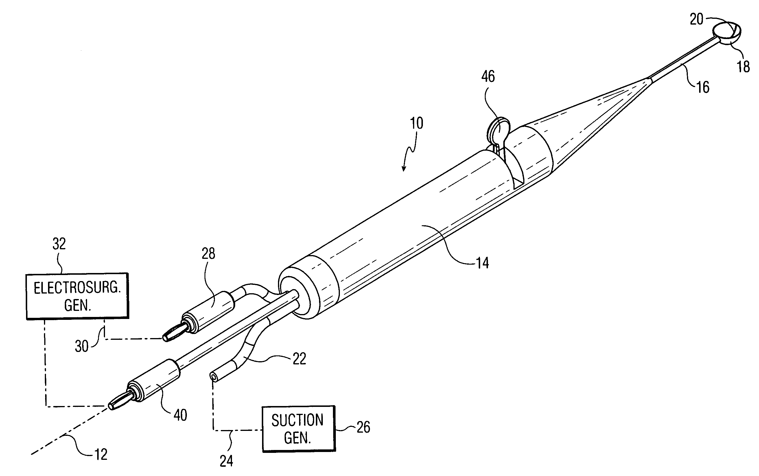
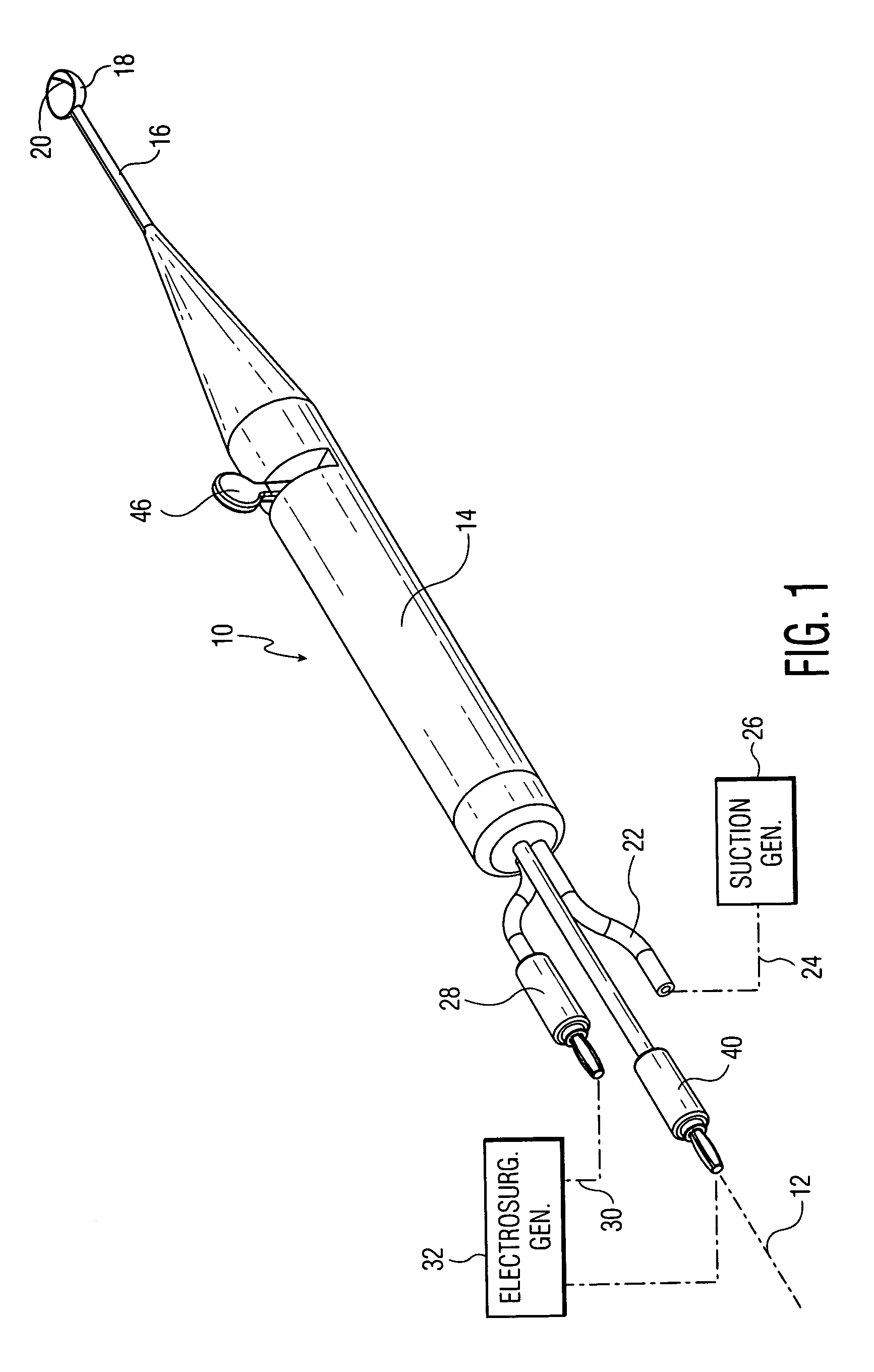
[0039]FIGS. 6–10 illustrate another embodiment 60 of the invention wherein the tissue-clearing member is a rotary vane. In this embodiment, it is preferred that the cup have a cylindrical shape. In the perspective view of FIG. 6, the non-working end at the left of the drawing is similar to that in the FIG. 1 embodiment, with a suction tube 22, and two electrical connectors 28, 40 extending out of the back of the tubular housing 62 that serves as the instrument handle. There is also a thumb-activating lever 64 present in this embodiment that is operably connected to the working end at the right of the drawing. The working end in this instance comprises a cylindrical scoop- or cup-shaped member 66 along the center axis of which is mounted an axle 68 connected to a radially-extending vane or element 70. The mounting is such that the radial element can rotate in a plane parallel to the open face 72 of the cup together with its axle 68, when the latter is rotated. As before, one electrod...

first embodiment

[0042]In operation, the surgeon manipulates the handle 60 with the leading edge loop 74 active for unipolar operation while pressing the leading edge into the spinal tissue and forcing the tissue into the cup 66, the tissue in the cup being removed via the suction connection 22 and interior channel 55, whose opening into the cup is shown at 58. Whenever deemed necessary, the surgeon can operate the tissue removing lever 64 to cut off the tissue inside the cup from that outside the cup allowing the suction room to perform its extraction function. Alternatively, the surgeon can electrically activate the tissue-clearing vane 70 during this excising action or while the cup is being pressed into the tissue to obtain bipolar operation. As with the first embodiment, the instrument can be constructed with just the loop 74, or with just the vane 70, or with both as illustrated for the most versatility in operation. To ensure that the two electrodes remain electrically-insulated, it is prefer...


PUM


Abstract

An electrosurgical instrument for spinal procedures comprises a generally scoop-shaped cup whose periphery is electrically active and is capable of applying RF electrosurgical currents to spinal tissue. The active electrode may comprise an exposed bare wire at the leading edge of the cup. A conduit in a handle section can convey suction to the cup. A tissue clearing member is pivotably mounted on or inside the cup and can be manipulated via a lever on the handle to help dislodge tissue. The tissue clearing member may be a radially-extending vane for rotation in the plane of the cup edge. The tissue clearing member may also be made electrically active thus selectively providing unipolar or bipolar operation.

Description

BACKGROUND OF THE INVENTION[0001]Spine fusion is the current gold standard of care for low back pain today. Spinal devices and procedures currently available do not always completely eliminate the source of a patient's pain or restore the patient's natural disc function or range of motion. There is also some evidence that shows that fusion at a particular level may cause further degeneration to the discs at adjacent levels. Spinal Arthroplasty nowadays is becoming coming more popular as it potentially can significantly reduce pain, restore nearly full range-of-motion, and reestablish natural disc height thereby keeping the facet joints and surrounding ligament structure and tissue intact. Artificial disc prostheses and disc augmentation techniques have been under development for over 35 years. The evolutionary course of artificial disc product development has included an array of product designs, materials and instruments. Mechanical, elastometric and physiological solutions have be...

Claims


Application Information

Patent Timeline
Patent Type & Authority Patents(United States)
IPC IPC(8): A61B18/18A61M1/00
CPCA61B17/32002A61B18/1402A61B2017/306A61B2018/0044A61B2018/0097A61B2218/007A61B18/12A61B18/00
Inventor GARITO, JON C.ELLMAN
Owner ELLIQUENCE
Learn More