
Method and apparatus for performing intra-operative angiography

a technology of intraoperative angiography and angiography, which is applied in the field of methods and apparatus for performing intraoperative angiography, can solve the problems of poor coronary artery function, poor blood circulation, stenosis and occlusion

Inactive Publication Date: 2005-07-05
STRYKER EUROPEAN OPERATIONS LIMITED
View PDF21 Cites 163 Cited by
  • Summary
  • Abstract
  • Description
  • Claims
  • Application Information

AI Technical Summary

Benefits of technology

[0014]A further aspect of the present invention permits a physician to accurately determine the extent to which a selected portion of body tissue, e.g., heart tissue, tumor, is well perfused, to assist in the identification and diagnosis of improperly (or properly) perfused tissue. The method comprises the steps of selecting a portion of body tissue to be analyzed; administering a fluorescent dye to the patient; obtaining at least one angiographic image of the selected tissue; and examining the at least one angiographic image to assess the extent of blood flow within the selected portion of body tissue.

Problems solved by technology

In coronary arteries, stenosis and occlusion often manifest themselves in the form of severe chest pains and, potentially, myocardial infarction (heart attack).
Poor patency in the first few months after surgery is thought to be the result of various factors, with the following believed to be the most significant: poor blood circulation, poor coronary arterial runoff, injury to the graft during preparation or faulty surgical technique.
While cardiac surgery in recent years has focused on strategies to minimize trauma to the myocardium, these strategies may increase the likelihood of problems if used during vessel grafting procedures.
However, this method has also raised concerns relating to graft quality.
Indeed, there exist reports of early failure in grafts completed using limited access incisions.
Other issues affecting CABG procedures are diagnostic in nature, and include relatively slow and inaccurate identification of stenotic and occluded vessels during the initial phase of CABG procedures (as some of these vessels lie within the heart tissue which inhibits visual identification), and an inability to quickly and accurately determine the extent of blood flow through the relatively smaller downstream vessels (and, more generally, whether the graft was successful in restoring blood flow to affected tissue) after the graft is completed.
Arterial patency issues may arise in therapies that do not include grafts.
While vessel patency information in these contexts may be obtained using X-ray technology, the disadvantages mentioned previously remain.
At present, MRI may be used to obtain perfusion information, but this information is imprecise and only available after treatment is completed.
Another affliction that requires treatment of the circulatory system is renal failure.
While X-ray technology can be used to assist the physician in determining whether the creation of a properly functioning fistula is possible, and the type of fistula that should be created, the technology suffers from the previously mentioned limitations.

Method used


Image

  • Method and apparatus for performing intra-operative angiography
  • Method and apparatus for performing intra-operative angiography

Examples


example

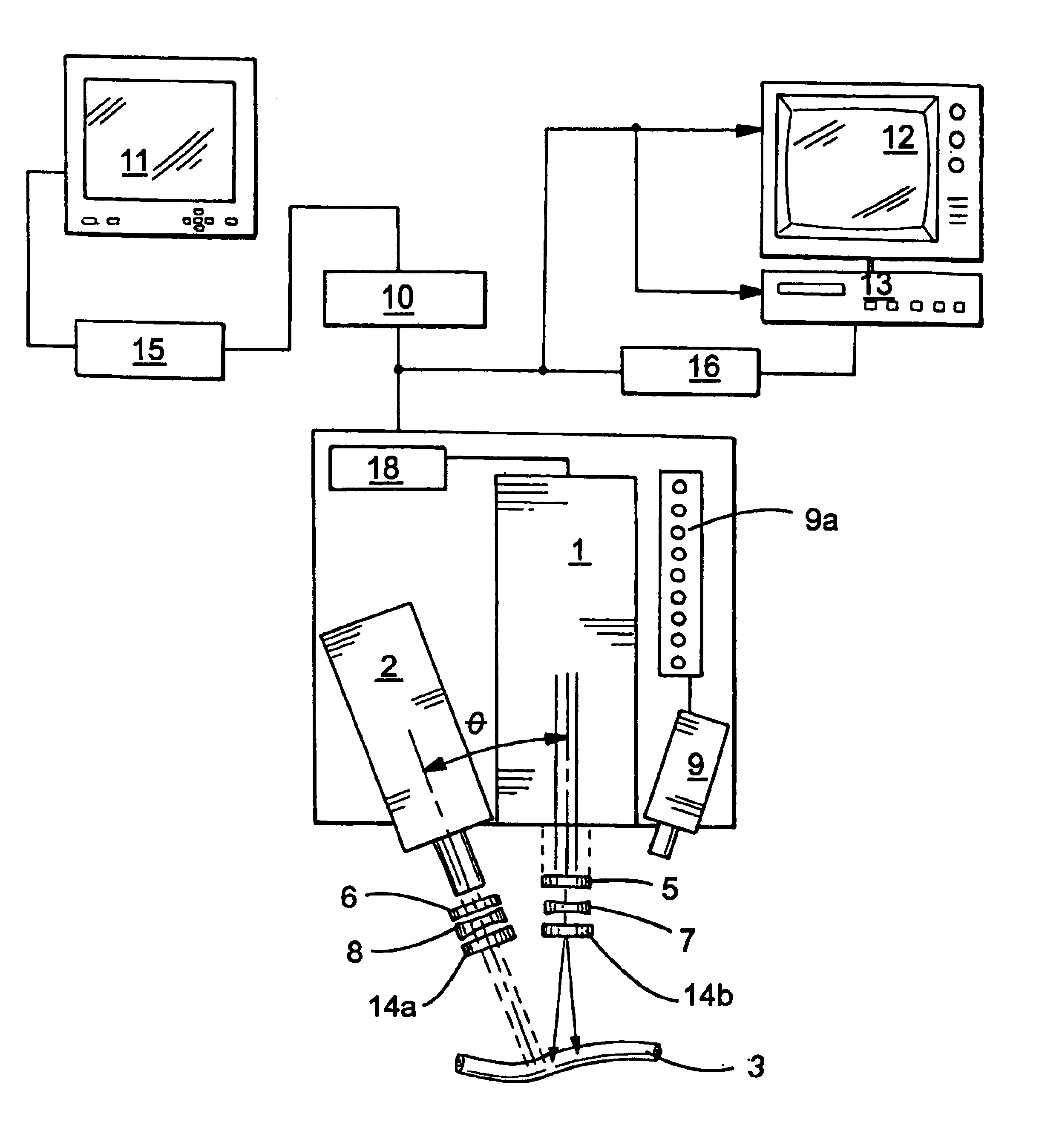
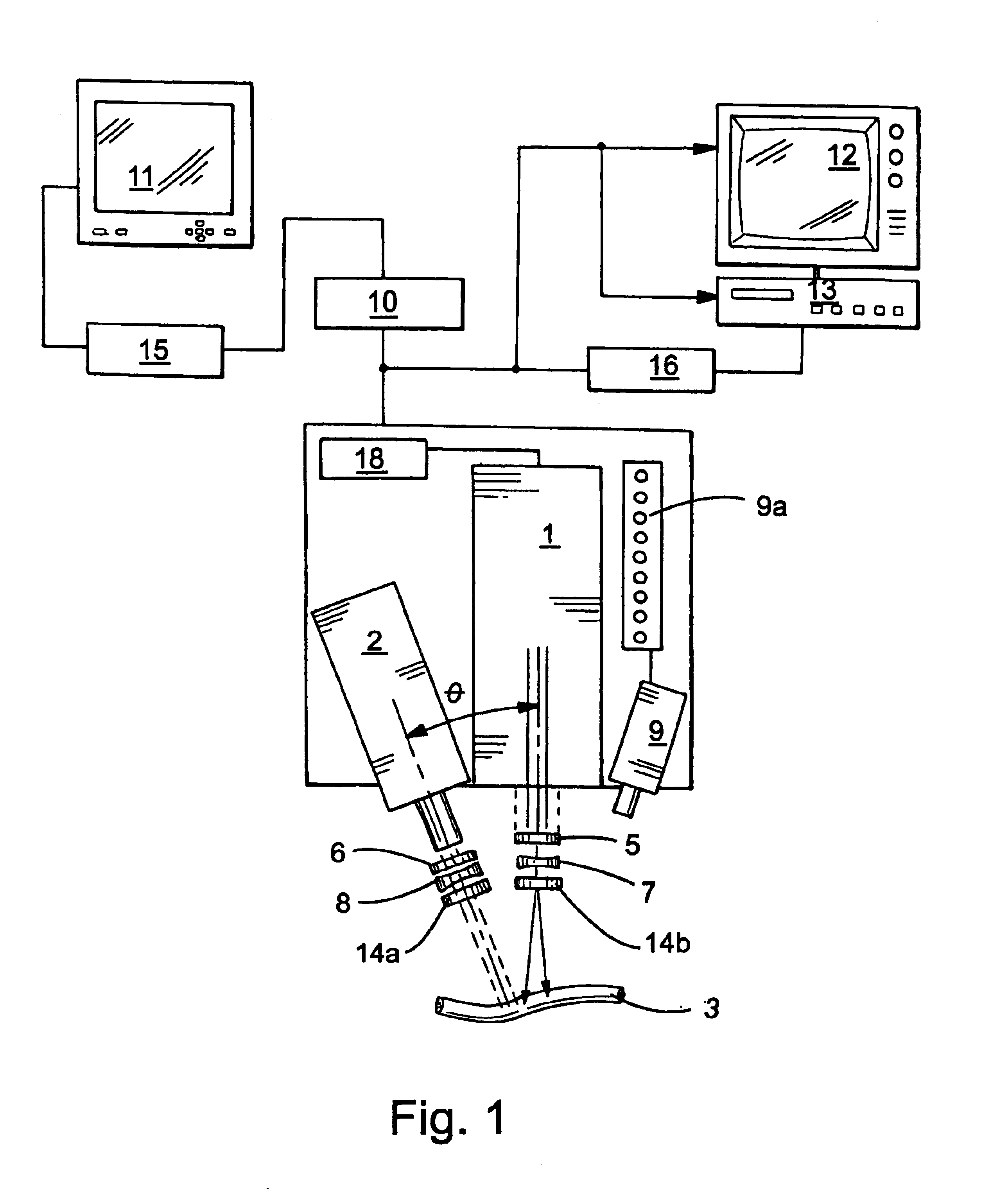
[0064]This example demonstrates the use of a preferred apparatus of the present invention in observing the flow of a fluorescent dye through a particular vessel, i.e., a mouse femoral artery, and langendorff perfused heart, and also demonstrates the ability of the apparatus to determine the diameter of a mouse femoral vessel under both normal conditions and under the influence of topically applied acetylcholine.

[0065]In this example, a fluorescent dye (ICG) was injected into the vascular bed (via jugular cannulation in the mouse: via an infusion line in the langendorff perfused heart) and excited using radiation from a laser source (806 nm). The fluorescence (radiation) emitted by the dye (830 nm) was captured as a series of angiograms using a CCD camera. The camera relayed the angiograms to analog-to-digital conversion software running on a PC that digitized the angiograms. The digitized images were then analyzed both qualitatively (by viewing the monitor) and quantitatively. One e...


PUM


Abstract

Method for assessing the patency of a patient's blood vessel, advantageously during or after treatment of that vessel by an invasive procedure, comprising administering a fluorescent dye to the patient; obtaining at least one angiographic image of the vessel portion; and evaluating the at least one angiographic image to assess the patency of the vessel portion. Other related methods are contemplated, including methods for assessing perfusion in selected body tissue, methods for evaluating the potential of vessels for use in creation of AV fistulas, methods for determining the diameter of a vessel, and methods for locating a vessel located below the surface of a tissue.

Description

CROSS-REFERENCE TO RELATED PATENT APPLICATIONS[0001]This patent application claims the benefit of U.S. provisional patent application No. 60 / 155,652, filed Sep. 24, 1999.TECHNICAL FIELD OF THE INVENTION[0002]This invention generally pertains to procedures for observing blood flow through the cardiovascular system of an animal.BACKGROUND OF THE INVENTION[0003]Disease and injury affecting the cardiovascular system in animals, and particularly humans, are commonplace in today's society. One such disease is atherosclerosis. This disease is characterized by partial blockage (stenosis) of a blood vessel, typically by a narrowing of one or more arteries. In its most severe form, the vessel narrows to the point that it becomes completely blocked (occluded). In coronary arteries, stenosis and occlusion often manifest themselves in the form of severe chest pains and, potentially, myocardial infarction (heart attack). Not limited to coronary arteries, atherosclerosis can also affect the periph...

Claims


Application Information

Patent Timeline
IPC IPC(8): A61B5/0275A61B5/026
CPCA61B5/0261A61B5/0275
Inventor DOCHERTY, JOHN C.HEWKO, MARKMANGAT, GURPREETFLOWER, ROBERT W.CHARI, SESHADRI M.
Owner STRYKER EUROPEAN OPERATIONS LIMITED
Learn More