Eureka AIR delivers breakthrough ideas for toughest innovation challenges, trusted by R&D personnel around the world.

System and method for determining reentrant ventricular tachycardia isthmus location and shape for catheter ablation

Inactive Publication Date: 2005-01-25
THE TRUSTEES OF COLUMBIA UNIV IN THE CITY OF NEW YORK
View PDF1 Cites 170 Cited by
  • Summary
  • Abstract
  • Description
  • Claims
  • Application Information

AI Technical Summary

Problems solved by technology

During ventricular tachycardia, the heart beats rapidly which can be debilitating to the patient and cause such things as tiredness and even syncope (i.e. fainting).
This clinical problem usually follows a myocardial infarction (heart attack) and is caused by abnormal electrical conduction in the heart because the cells become damaged during the infarct.
Therefore, because of both the poor filling action and the poor pumping action, there is less blood delivered to the tissues.
This causes the maladies that the patient experiences.
However, it is sometimes difficult to locate the best target site, and also the precise surface area to which energy should be delivered is often unknown and presently must be done by trial and error.
Although this former method potentially represents a substantial improvement over existing methods, it is not always convenient and cannot be used in all cases.
If tachycardia cannot be initiated, the former method described in the U.S. Pat. No. 6,236,883 to Ciaccio et al will not work because the methodology requires signals obtained from the heart surface during ventricular tachycardia.
Furthermore, sometimes tachycardia can be initiated but there is poor hemodynamic tolerance, which means that the pumping of blood is so poor during the tachycardia that the doctor must terminate it so that the patient does not experience syncope.
Ablating other areas can increase the chance of patient morbidity, by damaging regions of the heart unnecessarily.
Treatment of reentrant ventricular tachycardia by catheter ablation methods is hampered by the difficulty in localizing the circuit, particularly when the circuit structure is complex, the tachycardia is short-lived, or when reentry is not inducible during electrophysiologic study [1].
At the border zone, both normal and abnormal (low-amplitude, fractionated, or wide-deflection) electrograms are present [2-5]; however, these abnormal electrograms can be present both within and away from the reentry circuit location and are therefore not a specific predictor of its position in the border zone.
Therefore, methods for detection and measurement of abnormal sinus rhythm activation characteristics are not presently sufficient for targeting reentry circuits for catheter ablation, although presence of abnormality suggests the proximity of arrhythmogenic substrate.

Method used


Image

  • System and method for determining reentrant ventricular tachycardia isthmus location and shape for catheter ablation
  • System and method for determining reentrant ventricular tachycardia isthmus location and shape for catheter ablation
  • System and method for determining reentrant ventricular tachycardia isthmus location and shape for catheter ablation

Examples


Embodiment Construction

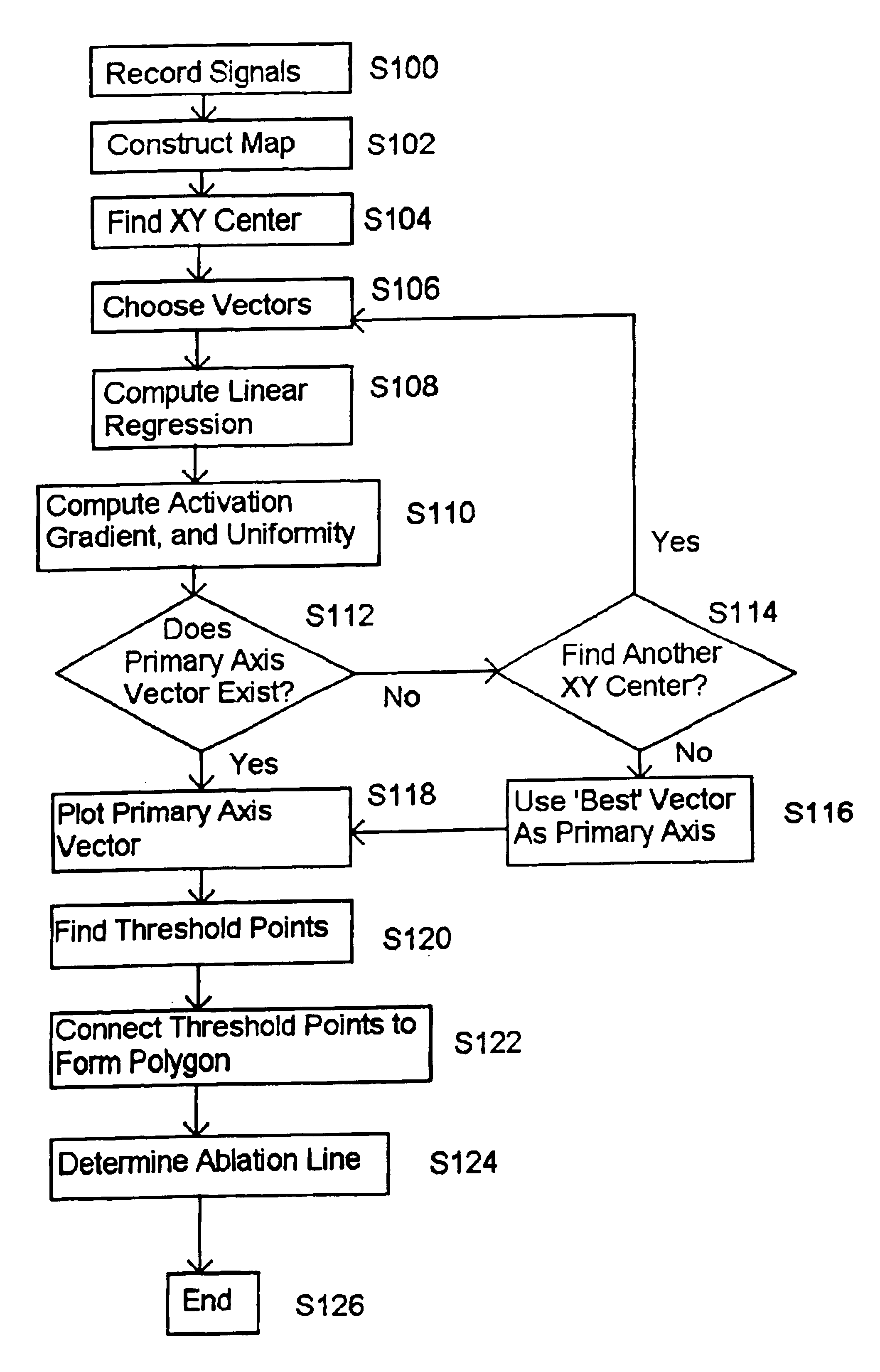
This invention provides a method for identifying and localizing a reentrant circuit isthmus in a heart of a subject during sinus rhythm, comprising the steps of: a) receiving electrogram signals from the heart during sinus rhythm via electrodes; b) creating a map based on the received electrogram signals; c) determining, based on the map, a location of the reentrant circuit isthmus in the heart; and d) displaying the location of the reentrant circuit isthmus.

In one embodiment of the above method, step b) includes arranging activation times of the received electrogram signals based on a position of the respective electrodes.

In one embodiment of the above method, the activation times are measured from a predetermined start time until reception of a predetermined electrogram signal.

In one embodiment of the above method, the map includes isochrones for identifying electrogram signals having activation times within a predetermined range.

In one embodiment of the above method, step c) incl...


PUM


Abstract

A method for identifying and localizing a reentrant circuit isthmus in a heart of a subject during sinus rhythm, including: a) receiving electrogram signals from the heart during sinus rhythm via electrodes; b) storing the electrogram signals; c) creating a map based on the electrogram signals; d) finding a center reference activation location on the map; e) defining measurement vectors originating from the center reference activation location; f) selecting from the measurement vectors a primary axis vector indicating a location of the reentrant circuit isthmus in the heart; g) finding threshold points of electrogram signals on the map; h) connecting the threshold points to form a polygon indicating a shape of the reentrant circuit isthmus in the heart.

Description

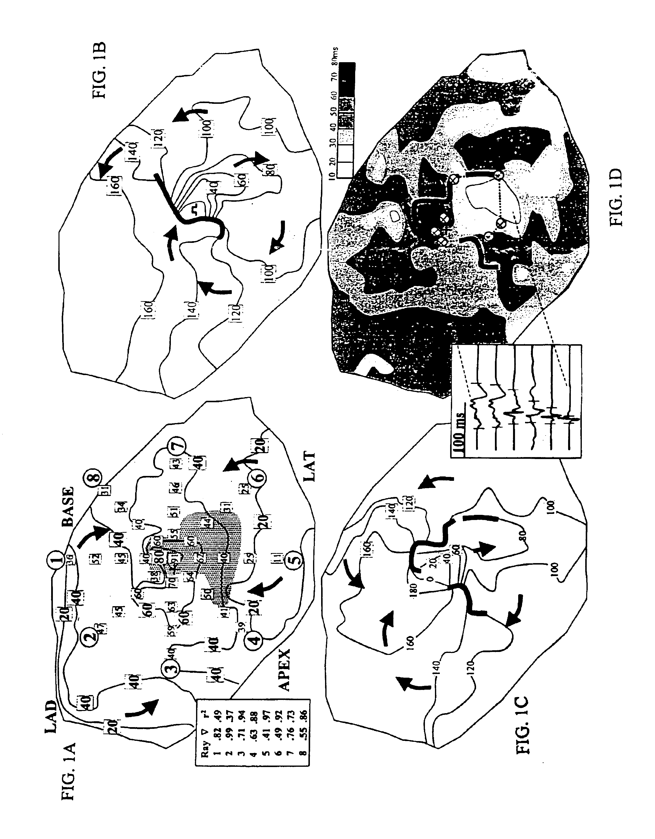
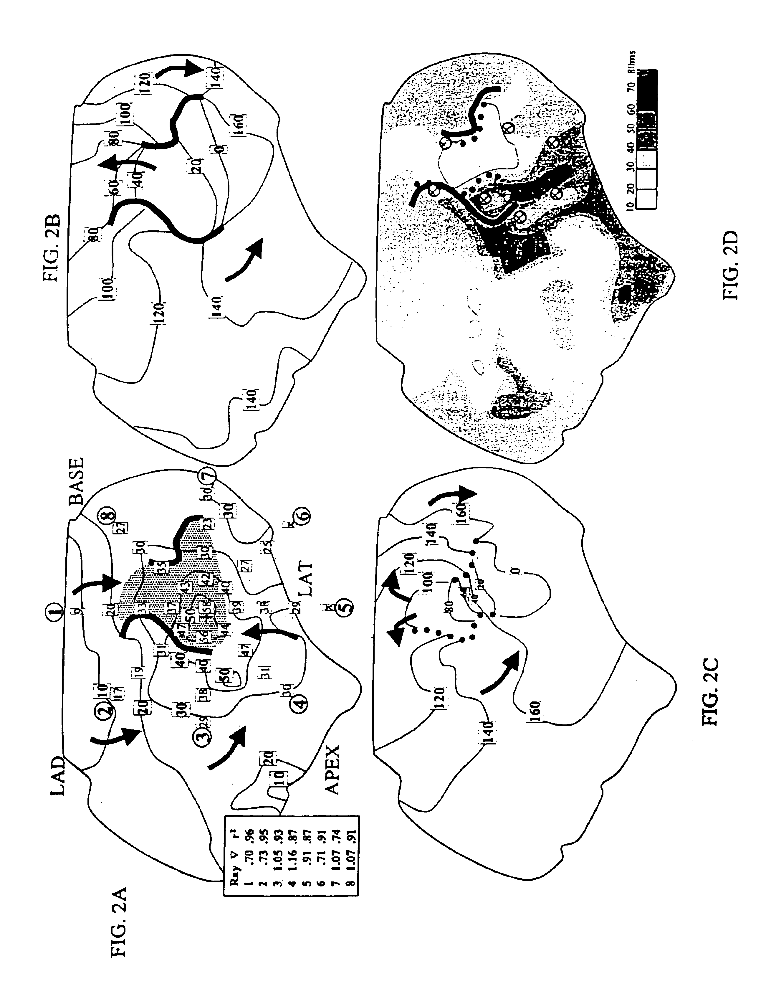
Throughout this disclosure, various publications may be referenced by Arabic numerals in brackets. Disclosures of these publications in their entireties are hereby incorporated by reference into this application to more fully describe the state of the art to which this disclosure pertains. Full citations of these publications may be found at the end of the specification.BACKGROUND OF THE INVENTIONIn canine hearts with inducible reentry, the isthmus tends to form along an axis from the area of last to first activity during sinus rhythm. It was hypothesized that this phenomenon could be quantified to predict reentry and the isthmus location. An in situ canine model of reentrant ventricular tachycardia occurring in the epicardial border zone was used in 54 experiments (25 canine hearts in which primarily long monomorphic runs of figure-8 reentry was inducible, 11 with short monomorphic or polymorphic runs, and 18 lacking inducible reentry). From the sinus rhythm activation map for each...

Claims


Application Information

Patent Timeline
IPC IPC(8): A61B5/0452A61B5/0402
CPCA61B5/04011A61B5/0452A61B18/1492G06F19/3437A61B2018/00357A61B2018/00839A61B2018/00898A61B2018/00214G16H50/50A61B5/341A61B5/349
Inventor CIACCIO, EDWARD J.WIT, ANDREW L.TOSTI, ALEXIS CHRISTINE
Owner THE TRUSTEES OF COLUMBIA UNIV IN THE CITY OF NEW YORK
Why Eureka
Eureka Blog
Learn More