
System and method of ultrasonic mammography

a technology of ultrasonic mammography and ultrasonic frequency, applied in the field of ultrasonic medical monitoring, can solve the problems of affecting the frequency in which the patient undergoes a mammography, and the access to the hospital or clinic, and achieve the effect of reducing the number of patients and reducing the number of procedures

Inactive Publication Date: 2002-11-19
MICROLIFE MEDICAL HOME SOLUTIONS
View PDF258 Cites 372 Cited by
  • Summary
  • Abstract
  • Description
  • Claims
  • Application Information

AI Technical Summary

Problems solved by technology

However, access to the hospital or clinic may affect the frequency in which the patient undergoes a mammography.
While the mammography devices currently used work well, some women may procrastinate in receiving a mammography, since the current method for obtaining an ultrasonic image of a breast typically often involves some discomfort to the patient, e.g. through pressing the breast as flat as possible.

Method used


Image

  • System and method of ultrasonic mammography
  • System and method of ultrasonic mammography
  • System and method of ultrasonic mammography

Examples


Embodiment Construction

)

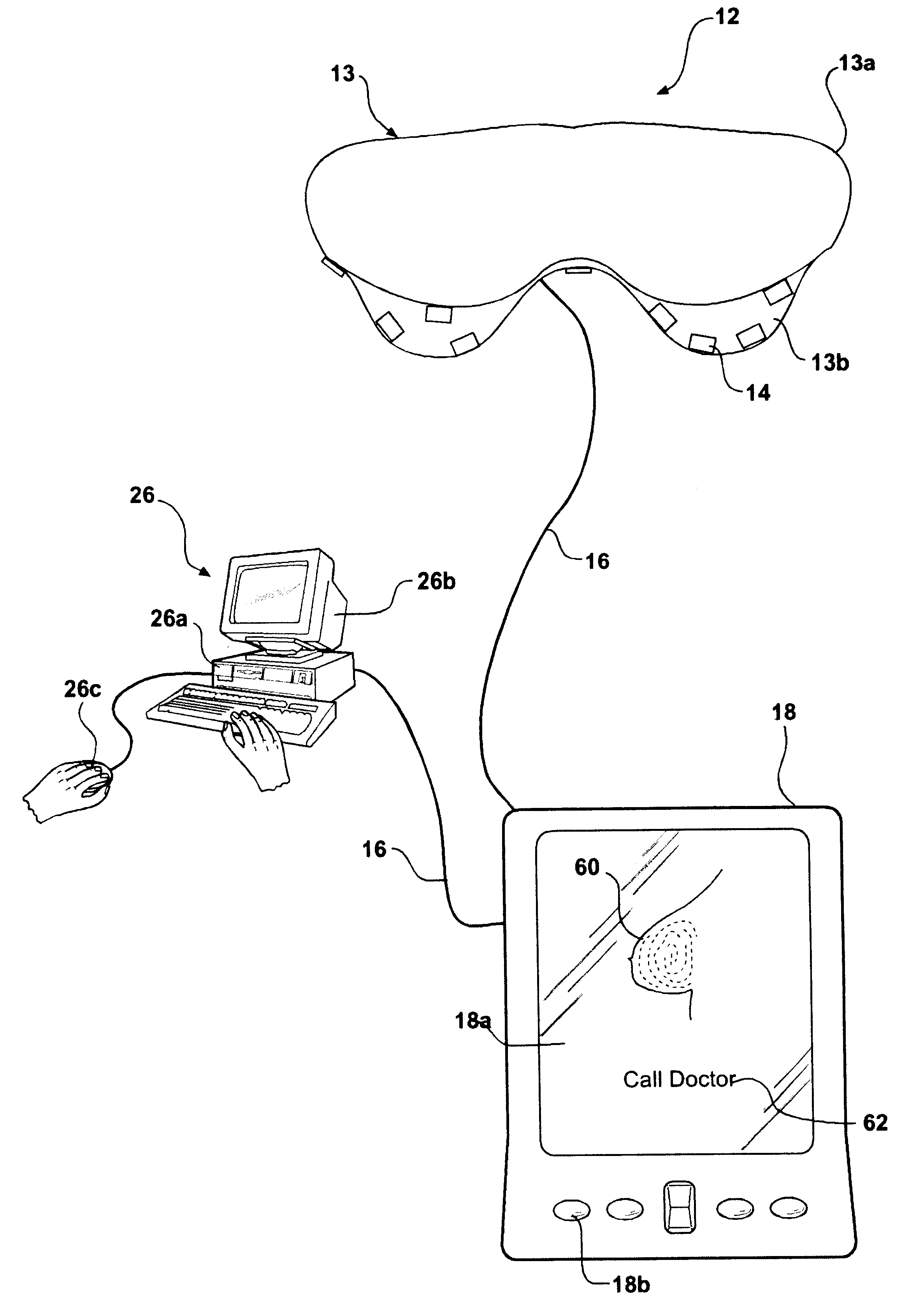
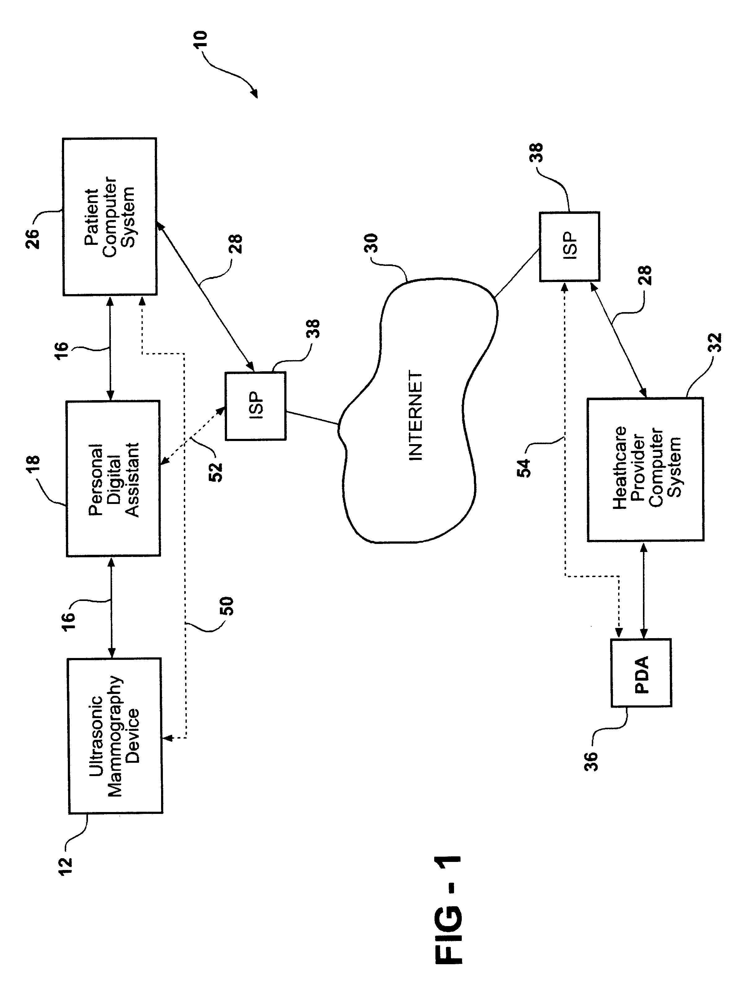
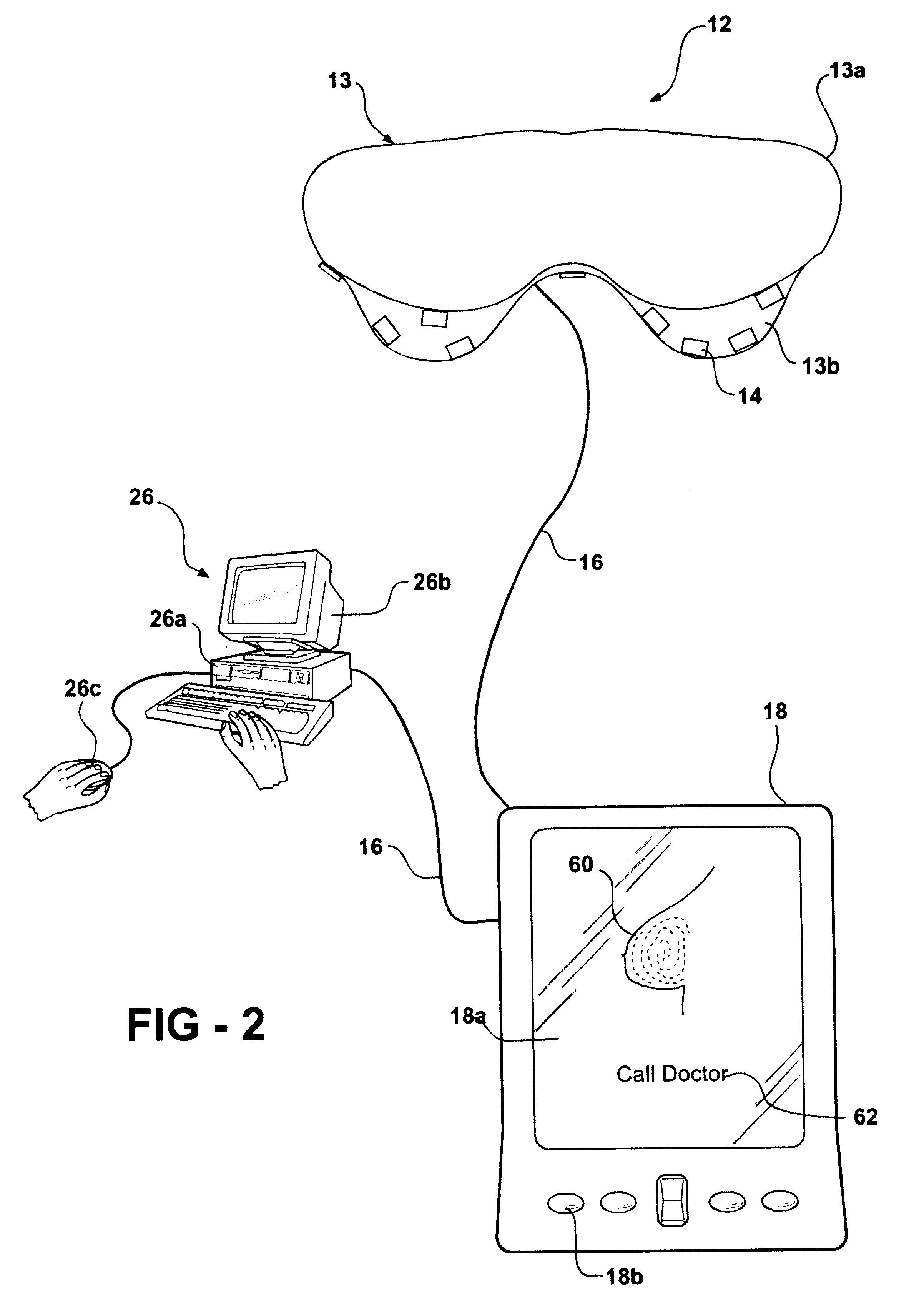
Referring to FIG. 1 a system for ultrasonic mammography is illustrated. The system 10 includes an ultrasonic mammography device 12 having a brassiere shape. The device 12 is intended to be worn by a patient (not shown) to obtain an ultrasonic image of the patient's breast. The ultrasonic mammography device 12 includes a support structure 13 having a frame portion 13a and a cup portion 13b attached to the frame portion 13a. It should be appreciated that the frame portion 13a and cup portion 13b are integral and formed as one piece. Preferably, the size of the frame portion 13a and cup portion 13b is adjustable, to accommodate a variety of patient sizes. It should be appreciated that a uniform medium, such as water, other liquids, a gel, etc. may be used to take up volume in the cups 13b not filled by the patient's breasts.

The ultrasonic mammography device 12 also includes a predetermined number of ultrasonic transducers 14 arranged in a predetermined manner on the support structure,...


PUM


Abstract

A system for ultrasonic mammography includes an ultrasonic mammography device for constructing an ultrasonic image of a breast having a support structure with an ultrasonic transducer mounted on the support structure. The system also includes a personal digital assistant operatively connected to the ultrasonic transducer via a communication link, a patient computer system operatively connected to the personal digital assistant via a second communication link, and a healthcare provider computer system operatively connected to the patient computer system via an internet, for constructing the image of the breast. The method includes the steps of positioning the ultrasonic mammography device having an ultrasonic transducer on the patient and activating the ultrasonic transducer to generate a signal for constructing an image of the breast. The method also includes the steps of transmitting the signal from the ultrasonic transducer to the personal digital assistant, and transmitting the signal via an internet to a healthcare provider computer. The method further includes the steps of using the signal to construct an image of the patient's breast.

Description

1. Field of the InventionThe present invention relates generally to ultrasonic medical monitoring and, more specifically, to a system and method of ultrasonic mammography for detecting a physiological condition of a patient's breast.2. Description of the Related ArtMedical monitoring involves the measurement of a physiological condition of a patient. An example of a medical monitor is disclosed in U.S. application Ser. No. 09 / 669,125 (filed Sep. 25, 2000) and Ser. No. 09 / 821,417 (filed Mar. 29, 2001, entitled "Monitoring System"), the disclosures of which are incorporated herein by reference. Conventional breast imaging is typically accomplished using a mammography ultrasound device within a medical setting, such as a hospital or clinic. The American Medical Association has set forth guidelines recommending how frequently a woman should undergo a mammography, since early detection of a medical condition, such as breast cancer, greatly affects the success of treatment for the conditi...

Claims


Application Information

Patent Timeline
Patent Type & Authority Patents(United States)
IPC IPC(8): A61B5/044A61B5/087A61B5/08A61B5/053A61B5/0402A61B5/22A61B5/00A61B5/083A61B5/11A61B5/024A61B5/022A61B7/00A61B8/08
CPCA61B5/044A61B5/0537A61B5/0833A61B5/087A61B5/222A61B5/4872A61B5/6817A61B5/6826A61B5/6838A61B5/6896A61B5/742A61B8/0825A61B5/0008A61B5/022A61B5/02438A61B5/14532A61B5/1455A61B7/00A61B2560/0295A61B2560/0456A61B2560/0462A61B2560/0468A61B2560/0475Y10S128/915A61B5/339
Inventor MAULT, JAMES R.
Owner MICROLIFE MEDICAL HOME SOLUTIONS
Learn More