Multilayer device for separating blood components and uses thereof

a multi-layer device and blood component technology, applied in the field of fluid sample component separation, can solve the problems of inaccuracy bias at the low and high ends of the hematocrit, many challenges in the product and testing method, and limitation of the processing of the collected sampl

Pending Publication Date: 2022-08-25
PARTNERSHIP FOR CLEAN COMPETITION
View PDF3 Cites 0 Cited by
  • Summary
  • Abstract
  • Description
  • Claims
  • Application Information

AI Technical Summary

Benefits of technology

[0007]In one embodiment, the invention provides a multilayer device for collection and separation of, for example, blood components, allowing for the detection of analytes from a fluid sample applied to the multilayer device, for example whole blood. The multilayer device may be a dried spot card composed in a book-type form in one embodiment, where there is a top and bottom cover hinged on one edge forming a spine and sandwiching multiple layers of membranes and materials similar to the pages of a book. Alternatively, more than one or all of the edges are temporarily connected or coupled, and any or all of the layers of the multilayer device that are connected or coupled on one or more edges, may be detached, removed, or separated from each other and from the device. Separation of the fluid sample allows for subsequent individual analyses of the separated components.
[0008]Another aspect may be directed to a multilayer device comprising layers that may be separately removed or detached as desired. A multilayer device having a rectangular shape, may be temporarily attached or coupled on all edges, where any or all layers of the multilayer device may be detached or removed. For example, the edges of any or all of the layers may be perforated in such a manner that allows for the layer to be tom out or detached from the remaining components of the multilayer device. Alternatively, the layers may be tightly sandwiched between the top and bottom covers in such a manner that the fluid sample does not leak and each of the layers of the multilayer device remain in position until separated for removal.
[0029]The benefits of the multilayer device and techniques described here include simplified sample collection, reduced costs, simplified shipping and storage, and a gained significant interest in various fields (Tretzel, L. et al. Analytical Methods 2015, 7, 7596-7605; Sadones, N. et al. Bioanalysis 2014, 6, 2211-2227). The inventive multilayer device overcomes many challenges in the art including the hematocrit effect and sampling of whole blood instead of plasma (De Kesel, P. M. et al. Bioanalysis 2013, 5, 2023-2041).
[0030]In another aspect, a multilayer device is configured for facile and rapid detection analyses of analytes found in dried sample spots collected and separated by the multilayer device, such as but not limited to automated, high-throughput analyses. Therefore, the inventive multilayer device described here was developed to be compatible for a wide range of hematocrit levels (e.g., 25%-65%), a high plasma volume yield from a whole blood fluid sample, and separate analyses of multiple components of a single fluid sample.

Problems solved by technology

Although sampling is advantageously achieved by a finger or heel stick to obtain a minimal volume, easy transport and sample stability, there are still several obstacles.
The available products and testing methods have many challenges with respect to limitations in sample collection and processing, sample yields, hematocrit (Hct) compatibility, and spectrophotometric detection.
One post-collection analyses issue is the limitation of processing the collected sample, which involves manual manipulation and detection from a hole punch by manually removing the dried blood spot, punching a small portion of the dried blood spot (about 3 millimeters-6 millimeters), and eluting the smaller sample in solvent for standard analyses.
With the wax boundary of the auto DPS card, the excess filtered plasma is retained within the boundary resulting in inaccuracy biases at the low and high ends of the hematocrit.
However, the card structures and production of plasma in each card format are different and each has its own disadvantages.
As described above, the auto DPS card may not be accurate nor efficient.
Although the NOVIPLEX™ card does not require any external device for generation of plasma spots, it is not compatible for automated analysis.
As a result, the sample handling process is tedious requiring a pair of tweezers to remove a small 2-mm filter disk containing the sample and manually transferring the disk for further sample extraction processes.
The yield of plasma volume is unfortunately low, i.e., 0.100 μL plasma per μL blood.
The plasma yield from the NOVIPLEX™ card is about 2.5 μL, which is insufficient for most analyses.
The low quantity of plasma is not due to a low initial blood sample volume, rather it is due to a limited capacity of the plasma collection substrate.
Scaling up is not possible.

Method used


Image

  • Multilayer device for separating blood components and uses thereof
  • Multilayer device for separating blood components and uses thereof
  • Multilayer device for separating blood components and uses thereof

Examples


example 1

Construction of Book-Type Multilayer Device

[0150]To prepare the filtration membrane unit, for example, RBC filter disks, a iPOCDX™ membrane filter sheet was punched using HARRIS Uni-Core™ punch of 5 mm diameter (Amazon, USA). A bottom support layer comprising cutout disks of 5 mm diameter, which used the card stock material similar to the top cover or top support layer, were prepared using an OSBORNE arch punch (Amazon, USA). As shown in FIG. 1, after preparing each layer of the materials, the card was assembled by first placing a polyester hydrophobic membrane layer (4) on the inner side of the underside of a top cover card stock (2) followed by punching equally spaced four holes through the upper card stock or top cover and through the polyester hydrophobic membrane layer (4). The bottom elevated or raised supports (6) were made of two round disks of the cover support card stock material affixed on the inner surface of the bottom cover card stock (7) using an adhesive, such as for...

example 2

Automated Flow-Through Elution Coupled with on-Line SPE-LC-MS / MS Bioanalysis of Opioids and Stimulants in Blood

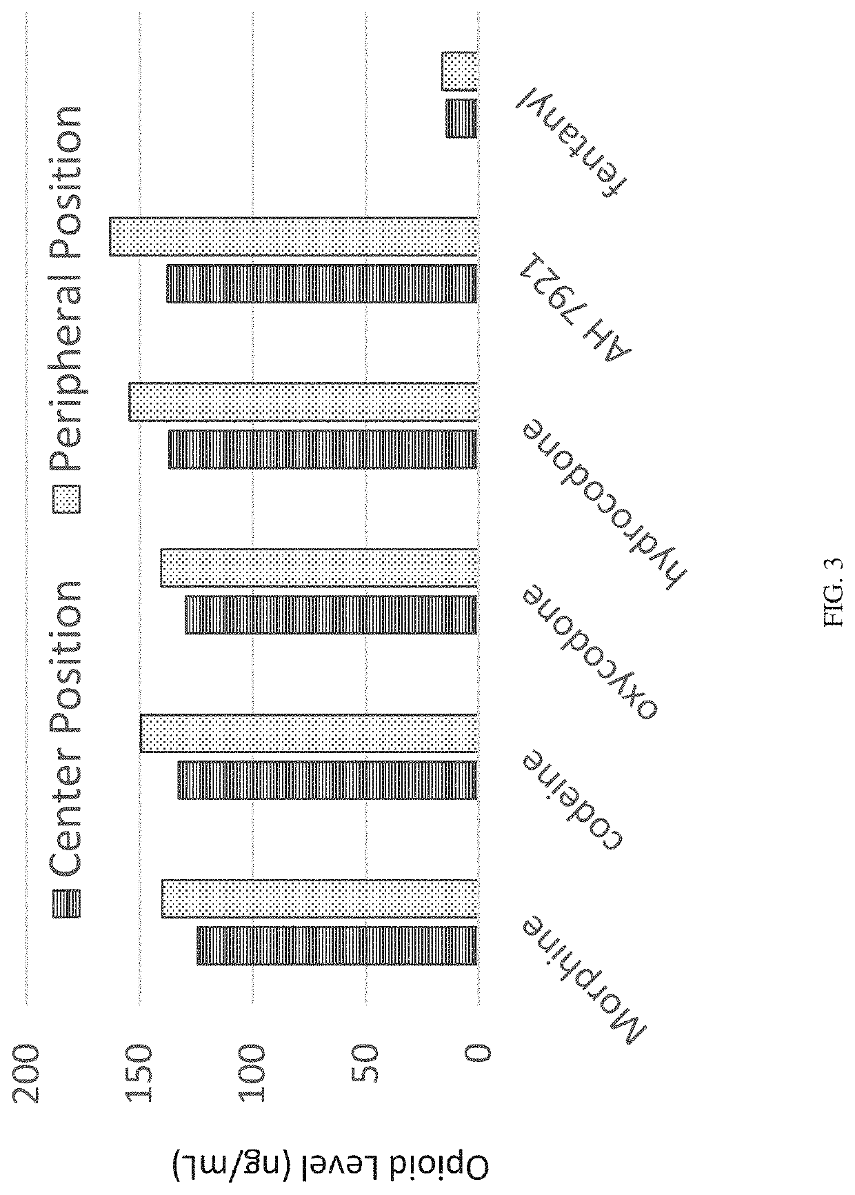
[0153]In order to design, develop, and validate a hematocrit-capable multilayer device that can produce plasma without the need for centrifugation that is suitable for automated on-line liquid chromatography with tandem mass spectrometry detection (LC / MS / MS) analysis, a multilayer device was developed to prepare dried plasma spot samples from whole blood microsampling. Extraction of the resulting dried plasma spot was accomplished by direct elution followed by an on-line solid phase extraction (SPE) and LC / MS / MS determination of analytes of interest, in this case, opioids and stimulants. Four opioids and five stimulants having varying physiochemical properties were selected to test in this analysis.

[0154]A series of standard working solutions were prepared by dilution of primary stock solutions with 3:7 methanol / water (v / v). Calibration standards were also prepared at 5 ng / ...

example 3

Test Compounds for Testing Multilayer Device

[0187]The multilayer device of the invention was tested using a known hypertension and ADHD drug, guanfacine, with its [13C, 15N3] internal standard. Guanfacine (C9H9Cl2N3O) has a monoisotopic mass of 245.0123 Da, while the internal standard [13C, 15N3]-guanfacine (13CC8H9Cl215N3O) has a monoisotopic mass of 249.0067 Da. These compounds were tested using a whole blood sample with a Blood:Plasma binding ratio (Ke / p) of 1.5 and analyzed using LC-MS / MS bioanalysis following the protocol described here.


PUM


Abstract

Embodiments of the invention are generally directed to analyte detection and products facilitating the collection, separation of sample components, and analyte detection. The multilayer device that allows for a rapid, easy, accurate, and efficient test of a fluid sample for analytes of interest and methods of collecting, separating components, and testing using the multilayer device are described in various embodiments of the invention.

Description

CROSS-REFERENCE TO RELATED APPLICATIONS[0001]This International PCT application claims the benefit of priority from U.S. Provisional Patent Application No. 62 / 299,226, filed Feb. 24, 2016, entitled, “Dried Plasma Spot Card For Automated Online Determination Of Opioids,” which is incorporated here by reference in its entirety.FIELD OF INVENTION[0002]This invention relates generally to separation of fluid sample components for analyte detection. More particularly, aspects of the invention are directed to a facile and accurate device for separation of various components of whole blood, including but not limited to red blood cells, white blood cells, platelets, and plasma, using a multilayer device or multilayer separation device for the separation of blood components and methods of using such device to detect analytes in the various blood components, for example, specifically on the cell surface and in the intra- and extra-cellular fluids, such as, but not limited to, chemical compound...

Claims


Application Information

Patent Timeline
Patent Type & Authority Applications(United States)
IPC IPC(8): G01N1/40G01N33/49G01N33/48B01L3/00G01N33/569
CPCG01N1/4077G01N33/491G01N2001/288B01L3/5023G01N33/56966G01N33/48G01N2001/4088B01L2300/0681B01L2300/0816B01L2300/165
Inventor HENION, JOHN DEGREERYONA, IMELDABOWERS, LARRY DONALD
Owner PARTNERSHIP FOR CLEAN COMPETITION
Why Eureka
Try Eureka