Eureka AIR delivers breakthrough ideas for toughest innovation challenges, trusted by R&D personnel around the world.

Methods and Apparatus for Detection and Imaging of Epileptogenicity from Scalp High-Frequency Oscillations

a high-frequency oscillation and epileptogenicity technology, applied in the field of methods and apparatus for detection and imaging of epileptogenicity from scalp high-frequency oscillations, can solve the problems of limited regional coverage, difficult to achieve, and difficult to achieve, and achieve the effect of facilitating clinical management of epilepsy

Pending Publication Date: 2021-04-15
CARNEGIE MELLON UNIV
View PDF4 Cites 2 Cited by
  • Summary
  • Abstract
  • Description
  • Claims
  • Application Information

AI Technical Summary

Benefits of technology

The present invention is a system and method for noninvasively detecting and measuring the tendency of a patient's brain to have epileptic seizures using electrophysiological source imaging (ESI). This method can help clinicians understand and manage epilepsy better by identifying the specific areas in the brain that are responsible for seizures and providing information for pre-surgical diagnosis and post-surgical evaluation.

Problems solved by technology

Patients with epilepsy are usually treated with anti-epileptic drugs (AEDs) to suppress or prevent seizures with frequent mild-to-severe adverse effects; however, about 30% of the epilepsy patients do not respond to drug treatments.
The last decades have observed numerous efforts in identifying the EZ, while it remains challenging even with all the combinations of diagnostic concepts and neurological modalities.
However, iEEG is an invasive procedure, prone to induce various risks such as cerebral hemorrhage and infection and is also limited to regional coverage.
However, scalp HFOs, though promising in revealing the underlying epileptogenicity, are impeded from clinical translation and application by several challenges.
First, current identification of HFOs in scalp EEG is mainly based on manual labeling, which is tedious and laborious, and it is difficult to detect these low-amplitude and brief events in noisy EEG recordings automatically.
Although there have been several automated detectors reported in iEEG studies, the development of such detectors is still deficient for noninvasive EEG recordings due to the high noise level and more artifacts in scalp EEG which might lead to low sensitivity or high false positive rates.
Second, there are insufficient evidences in the current detection techniques to distinguish pathological events from non-pathological events, especially from the physiological ones.
Therefore, simple measures, such as frequency and amplitude, might not serve well the purpose of correctly discriminating the pathological HFOs.
Stated differently, the presence of physiological high-frequency activities (HFAs), often sharing the same frequency band with the pathological HFOs, makes automatic distinction of clinically relevant HFOs complicated, which may even result in conflicting evidence of HFOs as a biomarker of epileptogenic brain.
Third, it is difficult to utilize HFOs in scalp EEG to monitor or delineate the underlying epileptogenicity due to the high noise level contaminating the high-frequency band which might lead ESI techniques to spurious cortical activities.
Up till now, only few studies demonstrated the potential approach to image HFOs via ESI in EEG or MEG localizing the EZ, however these studies are either limited to manual identification of HFOs or validated in a small scale, and most of them were only able to provide qualitative imaging results such as the congruence with clinical information instead of a quantitative estimation of the EZ.

Method used


Image

  • Methods and Apparatus for Detection and Imaging of Epileptogenicity from Scalp High-Frequency Oscillations
  • Methods and Apparatus for Detection and Imaging of Epileptogenicity from Scalp High-Frequency Oscillations
  • Methods and Apparatus for Detection and Imaging of Epileptogenicity from Scalp High-Frequency Oscillations

Examples


Embodiment Construction

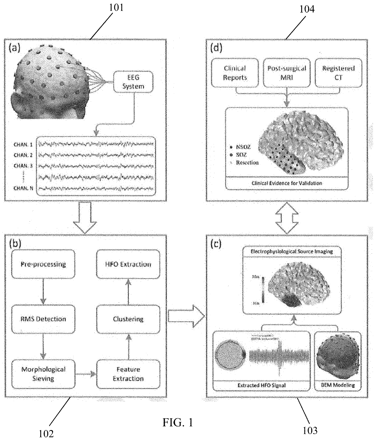
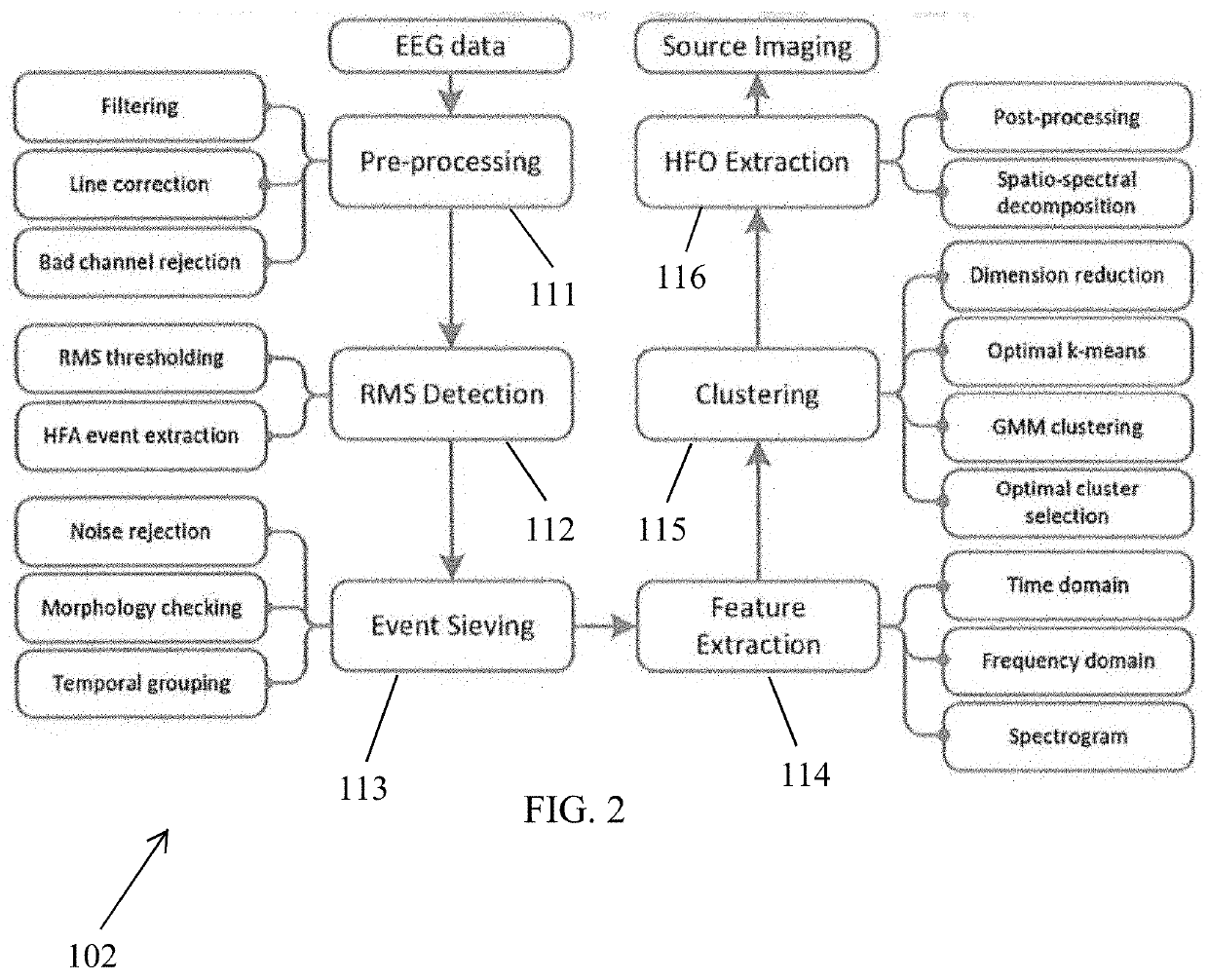
[0018]In epilepsy, high-frequency oscillations (HFOs) are observed and defined as events that stand out of the background with an approximately sinusoidal shape and a duration of at least four cycles. Studies have also observed nearly half of the ripples cooccurring with spikes (a typical form of inter-ictal epileptiform discharges, IEDs), termed “spike-ripples” (sRipples). These events are easier to detect than HFOs appearing alone and might be more related to pathological epileptic activities. Therefore, the method of the present invention, named Spike Ripple Source Imaging Algorithm (SPIRAL), can be used to detect, discriminate, and image the sRipples in scalp EEG for the purpose of delineating the epileptogenicity in epilepsy patients. A diagram of SPIRAL method is illustrated in FIG. 1.

[0019]The method consists of two major modules, namely, an identification module and an ESI module, which perform certain steps of the method. First, at step 101, scalp EEG data is recorded using...


PUM


Abstract

A system and method to facilitate the analysis of long-term scalp EEG recordings and the investigation of underlying epileptogenicity. Pathological high frequency oscillations are identified and extracted from the EEG recordings and electrophysiological source imaging is used to reconstruct cortical activity, which can be used as an aid in surgical resection for the treatment of epilepsy.

Description

CROSS-REFERENCE TO RELATED APPLICATIONS[0001]This application claims the benefit under 35 U.S.C. § 119 of Provisional Application Ser. No. 62 / 973,625, filed on Oct. 15, 2019, which is incorporated herein by reference.STATEMENT REGARDING FEDERALLY SPONSORED RESEARCH[0002]This invention was made with Government support under NS096761 awarded by the National Institutes of Health (NIH). The Government has certain rights in this invention.BACKGROUND OF THE INVENTION[0003]Epilepsy is one of the most common and severe chronic neurological disorder affecting approximately 65 million people in the world. Patients with epilepsy are usually treated with anti-epileptic drugs (AEDs) to suppress or prevent seizures with frequent mild-to-severe adverse effects; however, about 30% of the epilepsy patients do not respond to drug treatments. In such medically refractory epilepsy (MRE) patients, neurostimulation and resective surgery may be viable options to control or even stop seizures. Localizing t...

Claims


Application Information

Patent Timeline
IPC IPC(8): A61B5/04A61B5/00A61B5/0478
CPCA61B5/04012A61B5/4094A61B5/0478A61B5/6814A61B5/7207A61B5/7257A61B5/7246A61B5/7225A61B5/291A61B5/372A61B5/316
Inventor HE, BINCAI, ZHENGXIANG
Owner CARNEGIE MELLON UNIV
Why Eureka
Eureka Blog
Learn More