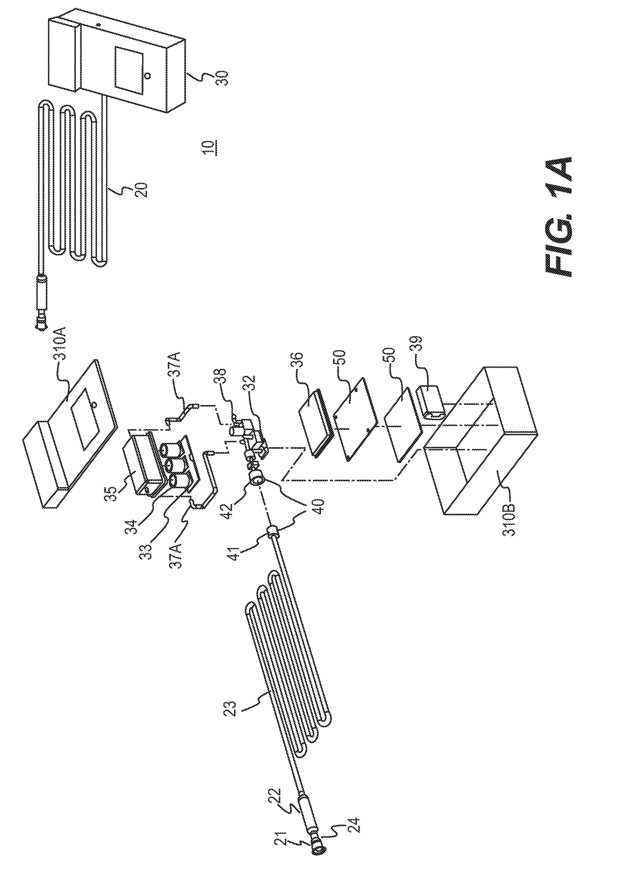
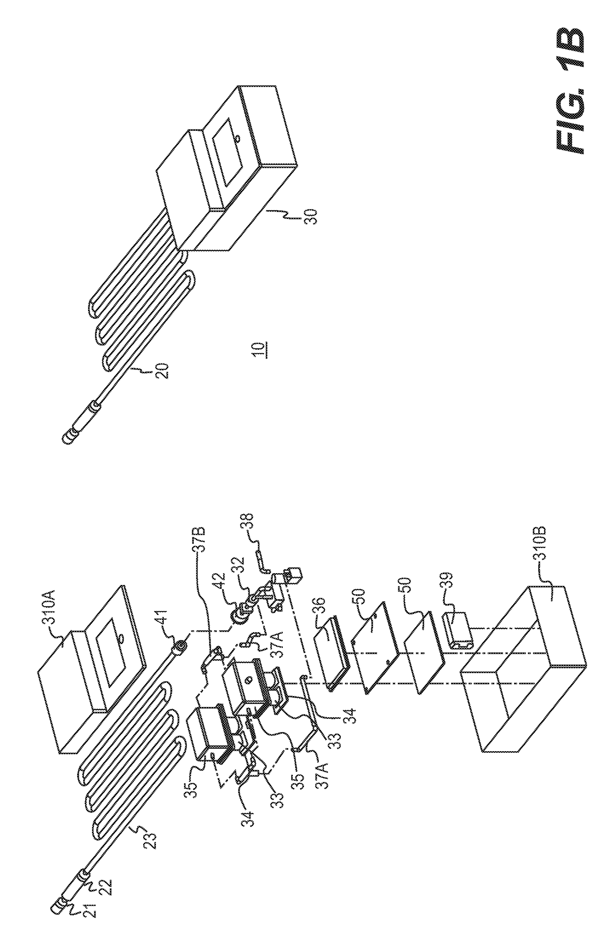
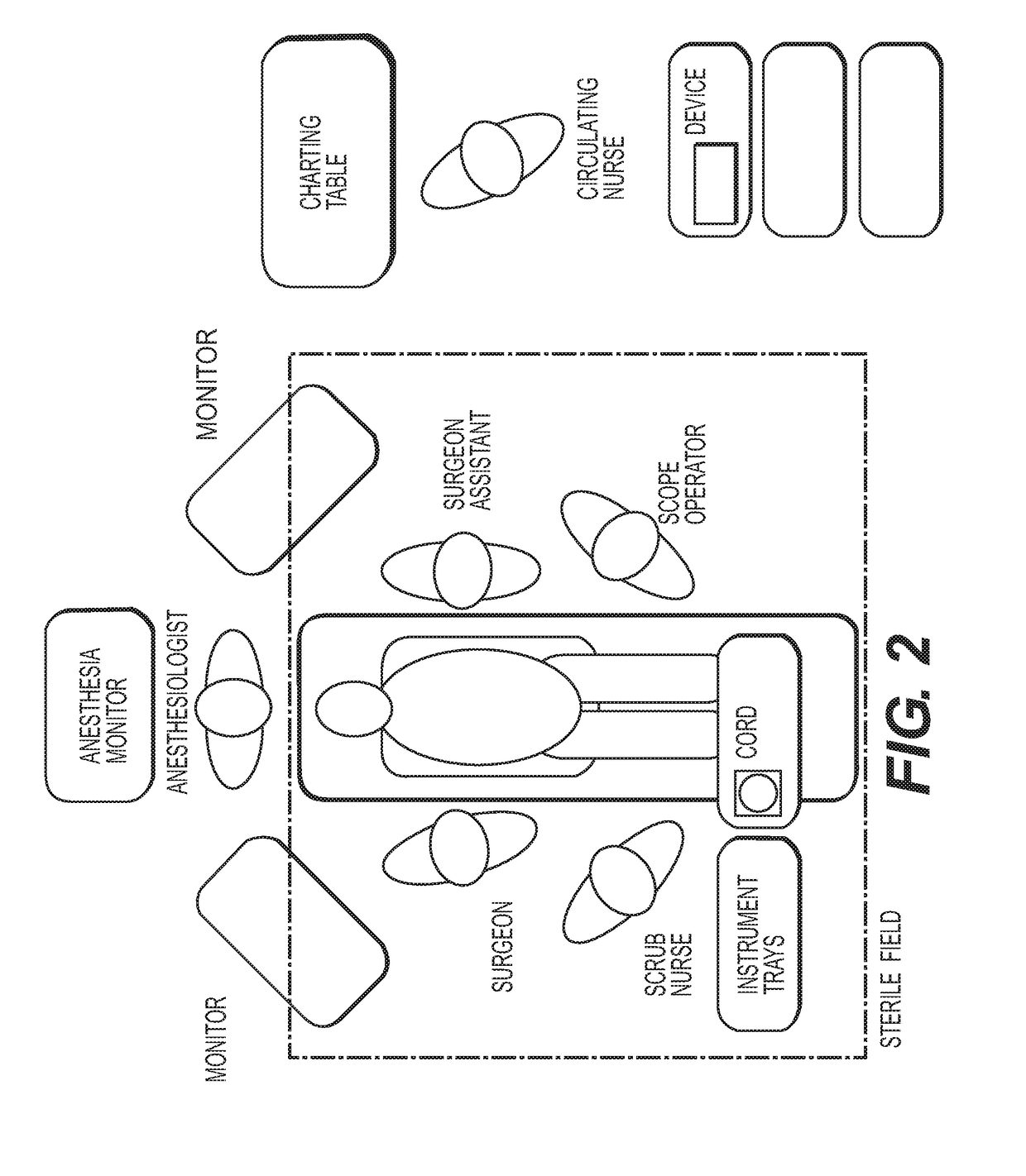
Methods and devices for detecting bowel perforation
a technology of bowel perforation and detection method, which is applied in the direction of separation process, instruments, applications, etc., can solve the problems of increasing the number and complexity of laparoscopic surgery that is routinely performed, the morbidity and mortality of patients is far greater, and the cost of reparation is increased
- Summary
- Abstract
- Description
- Claims
- Application Information
AI Technical Summary
Benefits of technology
Problems solved by technology
Method used
Image

Examples
example 1
[0152]Filter-Tubing Assembly Testing
[0153]A liquid bypass filter assembly is tested for the ability of the perforated tubing-absorbent sleeve assembly to achieve lateral fluid absorption in order to ensure a viable gas flow lumen. FIG. 29 is a photographic of the materials for use in the test. These include a Veress needle, colored water for visualization, a length of polyurethane medical grade tubing with a perforated section, a sheet of absorbent cellulose non-woven material, a rigid plastic housing, silicone sealant, and a syringe to serve as the pump. As shown in FIG. 30, the prototype bypass filter is constructed by inserting the perforated tube into the clear plastic housing. Following this, the absorbent cellulose wicking material is packed into the annular cavity created between the perforated section and the plastic housing. The ends of the plastic housing are sealed with silicone adhesive to prevent leakage from the annular space.
[0154]The testing process is shown in FIG. ...
PUM
 Abstract
Description
Claims
Application Information
 - R&D
- Intellectual Property
- Life Sciences
- Materials
- Tech Scout
- Unparalleled Data Quality
- Higher Quality Content
- 60% Fewer Hallucinations
Browse by: Latest US Patents, China's latest patents, Technical Efficacy Thesaurus, Application Domain, Technology Topic, Popular Technical Reports.
© 2025 PatSnap. All rights reserved.Legal|Privacy policy|Modern Slavery Act Transparency Statement|Sitemap|About US| Contact US: help@patsnap.com